
Scapula Bone Quiz
Hip Bone Anatomy
Last update:
Introduction
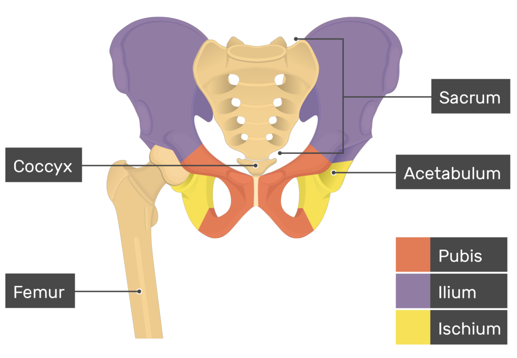
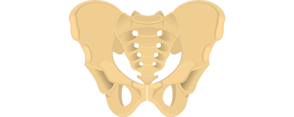
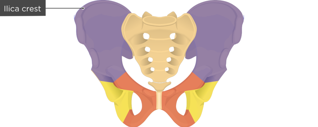
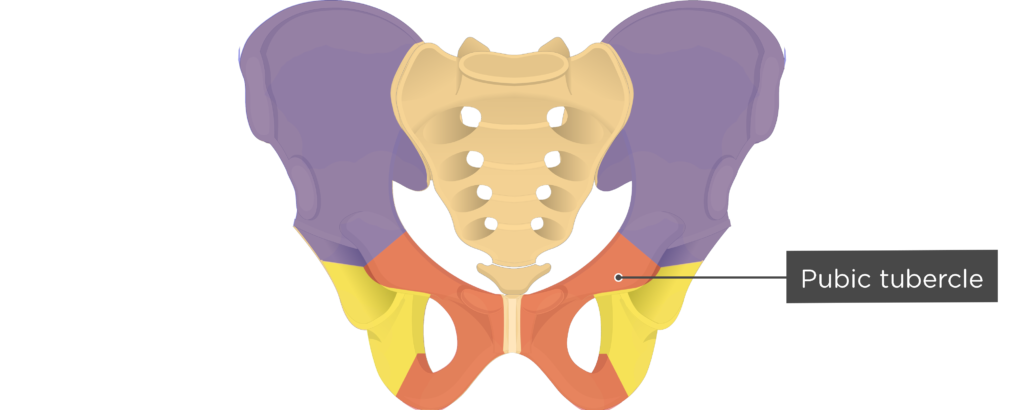
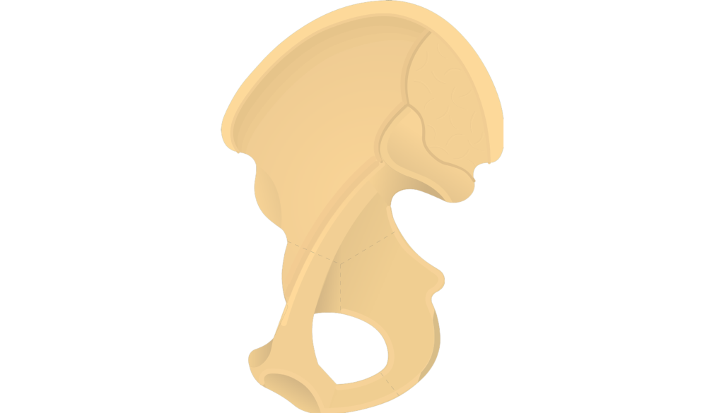
- Each os coxa bone (hip bone) is made up of
 three bones, which fuse during early adult-hood.
 - The ilium bone forms the superior portion of the os coxa, the ischium bone the lower posterior portion, and the pubic bone (pubis) the lower anterior portion.
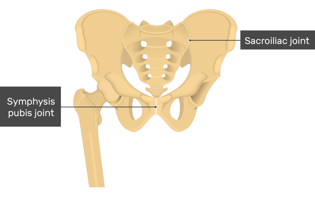
- Three articulation (joint) sites are found on each os coxa.
- The two os coxae meet anteriorly at the pubic symphysis joint and converge posteriorly with the sacrum at the sacroiliac joints. Laterally, deep sockets called acetabula accept the heads of the femurs to form the hip joints.
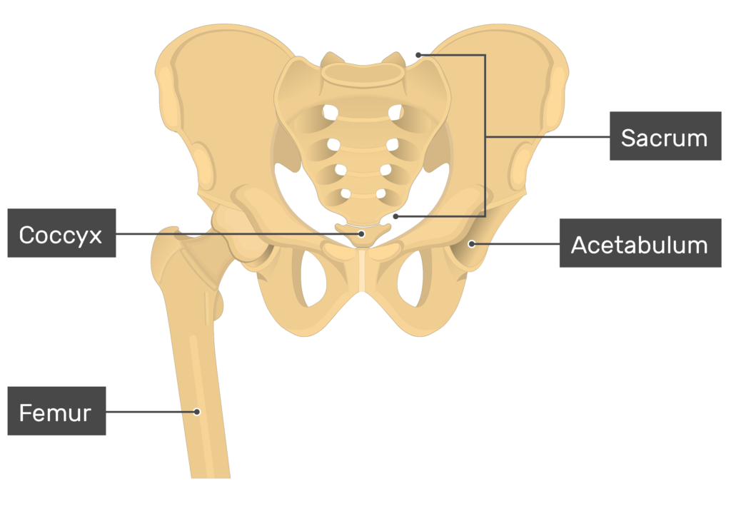

- Together, the right and left os coxae form the pelvic girdle. Together, the pelvic girdle, sacrum, and coccyx (final 3-5 vertebrae) make up a ring of bones called the pelvis (bony pelvis).
- The bony pelvis protects the soft organs of pelvic cavity (bladder, lower colon, rectum, and reproductive organs).
- It also provides attachment points for many muscles that control the movements of the back, abdomen, and femur bones.
Have you been making any of these common mistakes that hinder your anatomy learning?
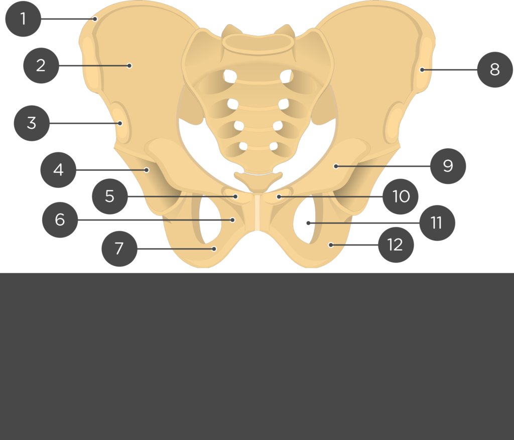
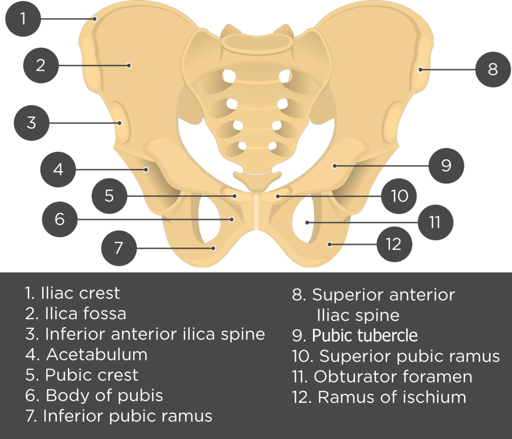
Anterior markings
Ilium Bone
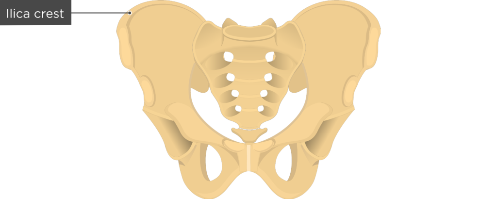
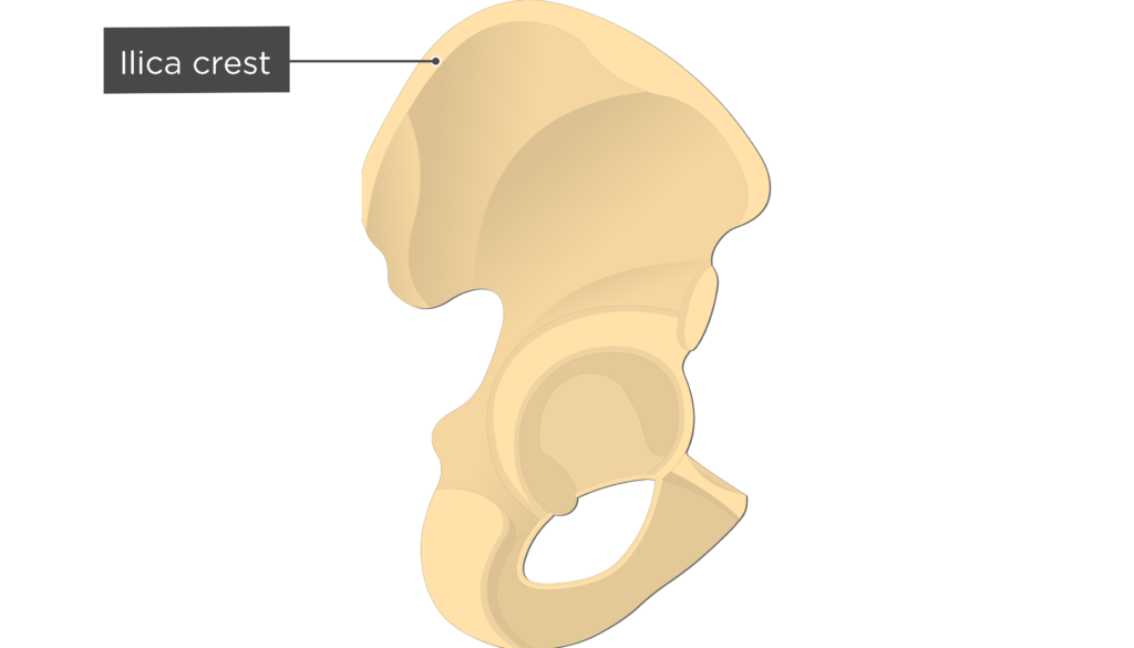
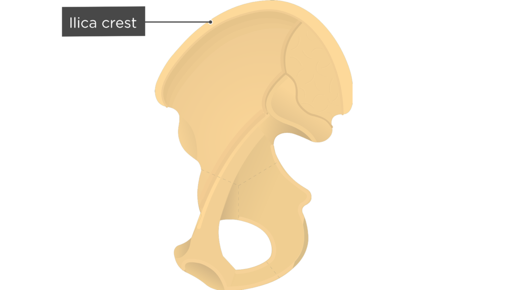
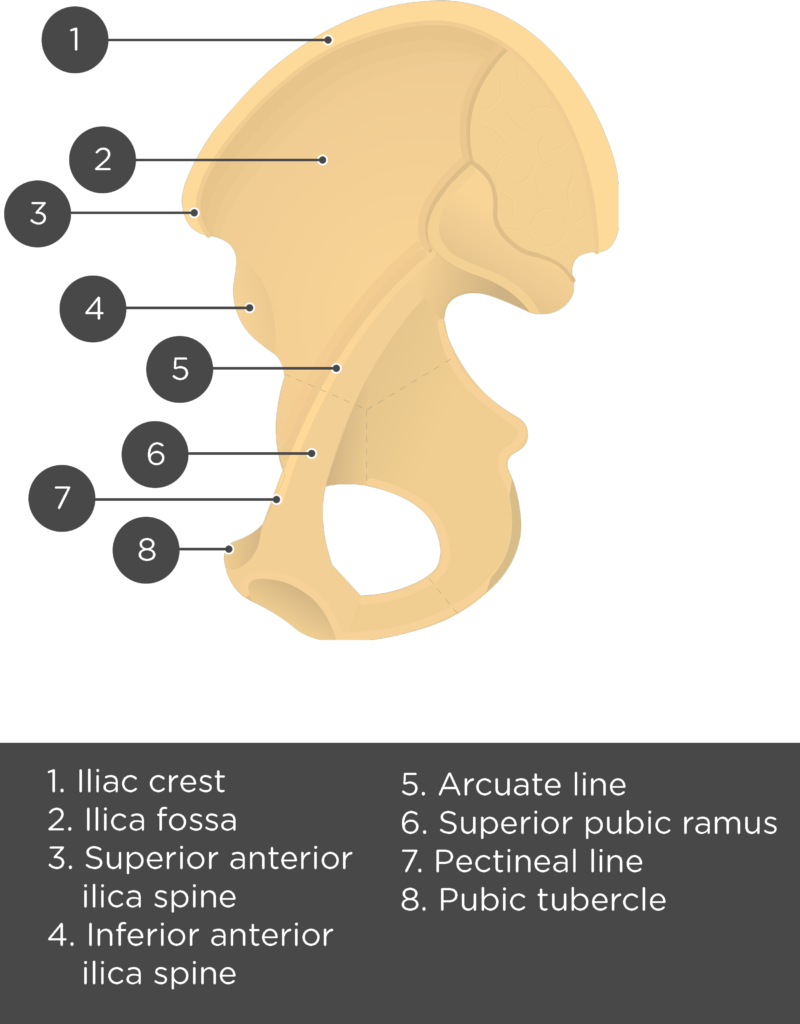
- Iliac Crest or Crest of Ilium (Crista iliaca) is the curved upper ridge of the ilium. The latissimus dorsi, quadratus lumborum, erector spine, iliacus, tensor fasciae latae, and abdominal muscles attach along the surface of this ridge.
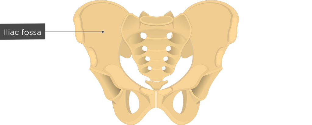
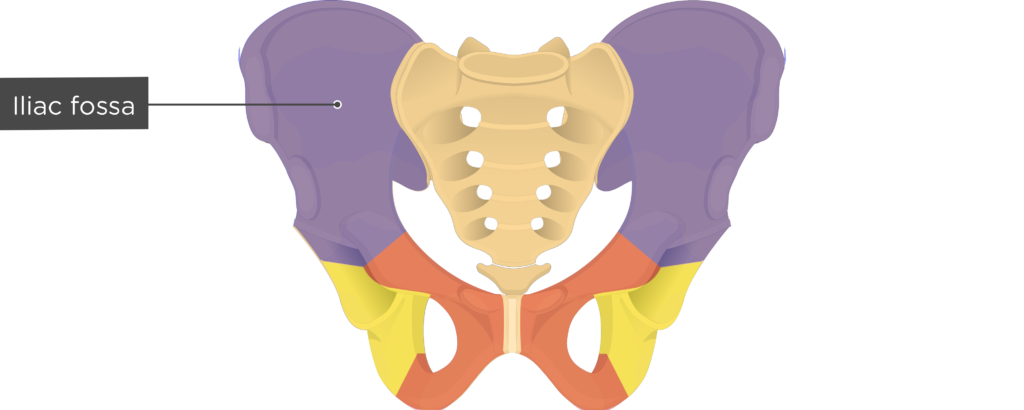
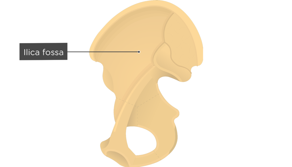
- Iliac Fossa (Fossa iliaca) is a broad depression located along the anteromedial surface, inferior to the iliac crest. It is an attachment point for the iliacus muscle.
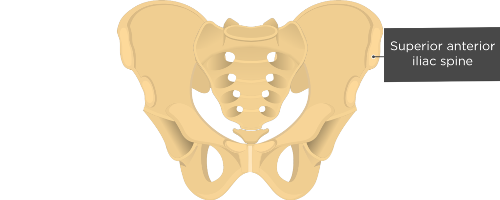
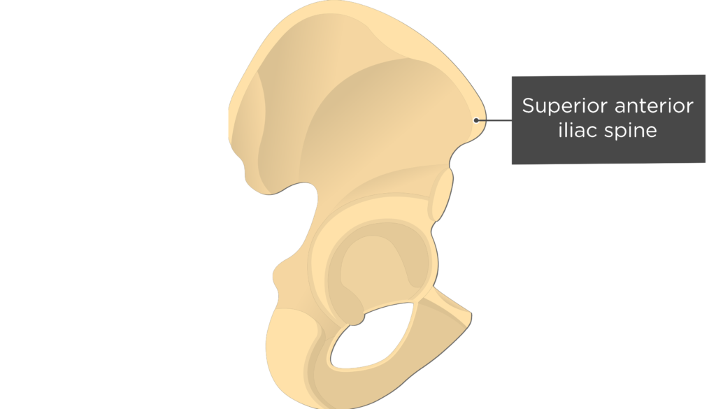
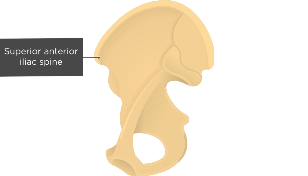
- Anterior Superior Iliac Spine (Spina iliaca anterior superior) is a anterior projection from the iliac crest that serves as an attachment point for the sartorius muscle and inguinal ligament.
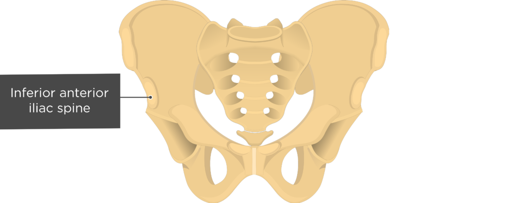
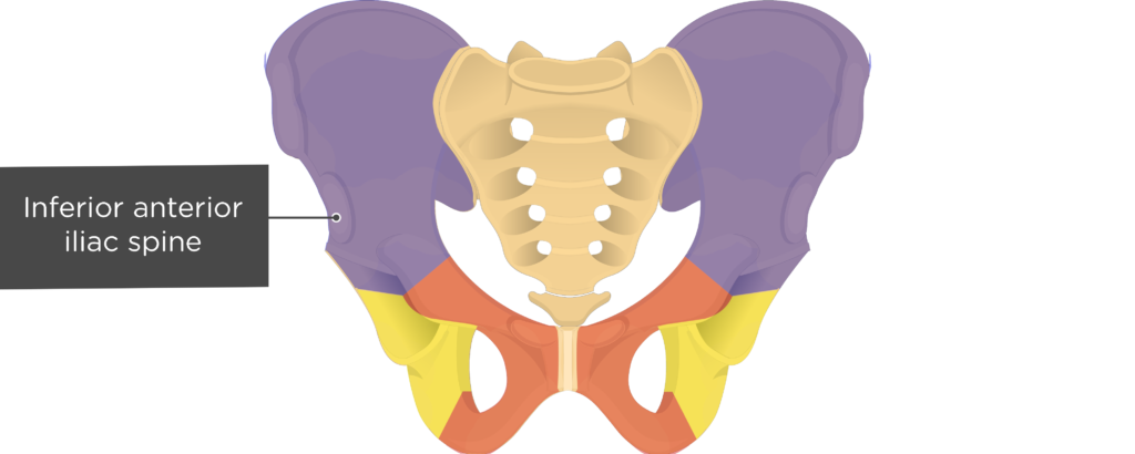
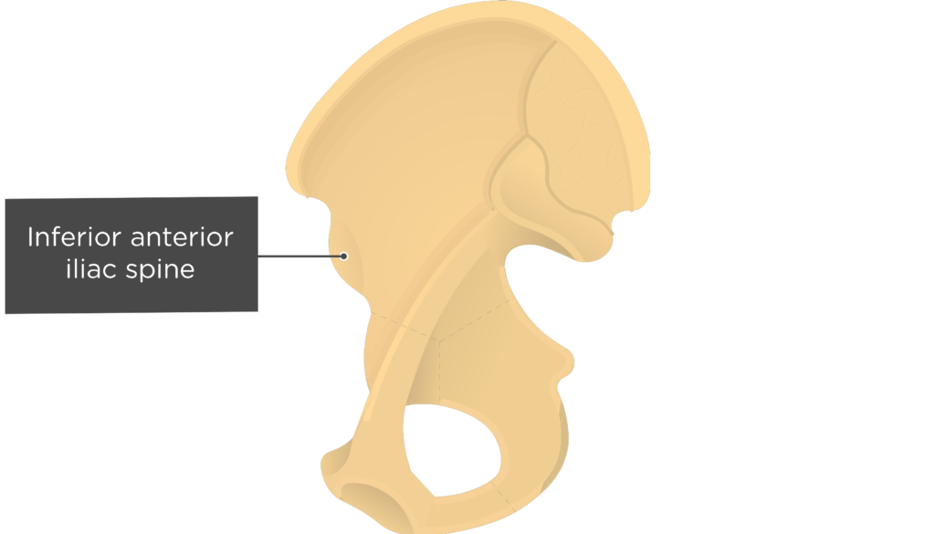
- Anterior Inferior Iliac Spine (Spina iliaca anterior inferior) is a projection below the anterior superior iliac spine that is as an attachment point for the rectus femoris muscle.

Pubic Bone (Pubis)
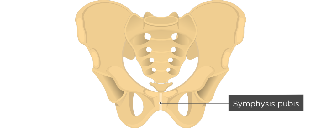
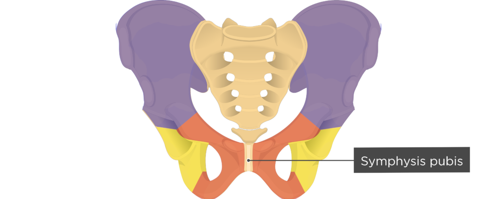
- Symphysis pubis or pubic symphysis (Symphysis pubica; symphysis pubis) is the cartilaginous joint between the two pubic bones.
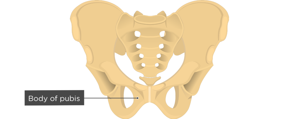
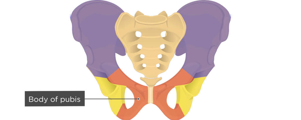
- Body of pubis (Corpus ossis pubis) is the flatten, medial end of the pubis that lies adjacent to the symphysis.
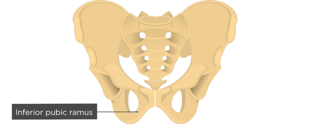
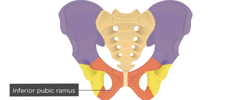
- Inferior Pubic Ramus (Ramus inferior ossis pubis) is a thin, flat, downward extension that runs diagonally from the medial pubis to the ischial ramus. The adductor brevis, adductor magnus, and gracilis muscles attach to this ramus.
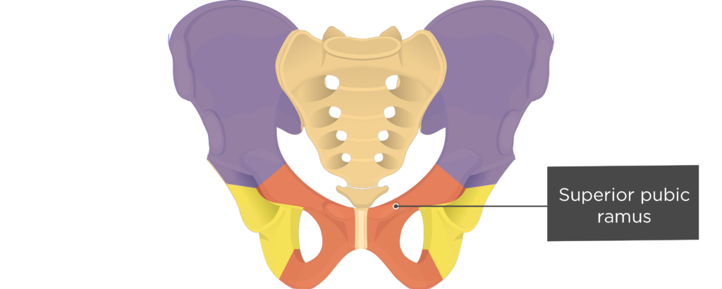
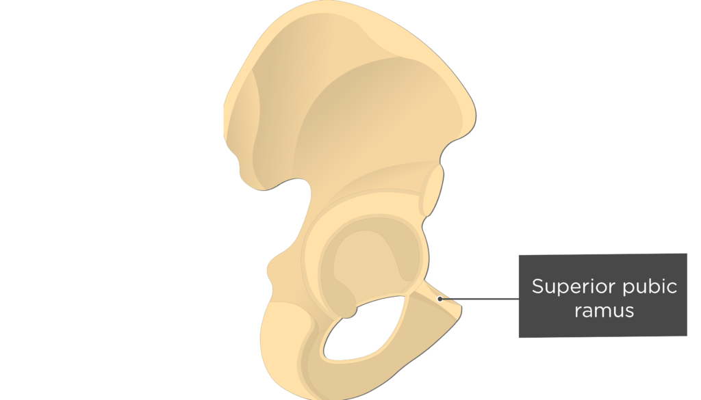
- Superior Pubic Ramus (Ramus superior ossis pubis) is a band of bone that runs along the superior aspect of the pubis. The surface of this ramus is an attachment site for the pectineus muscle.
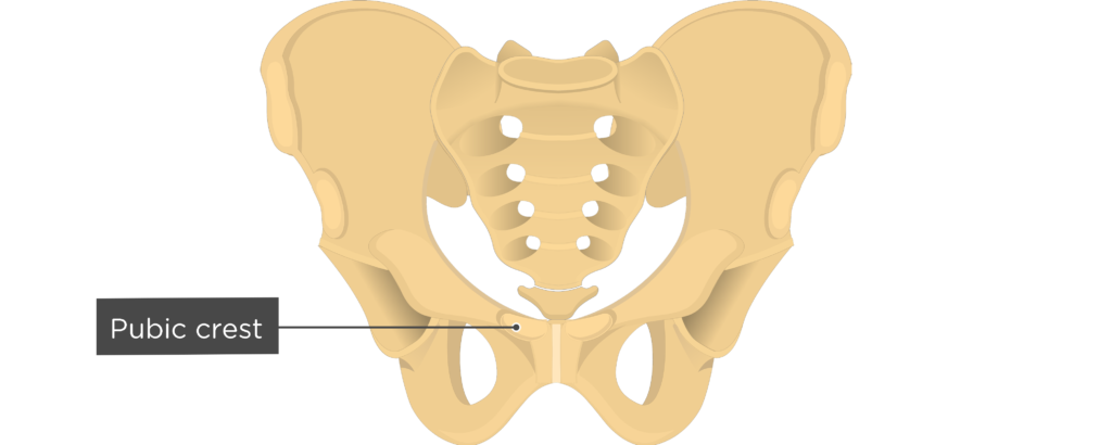
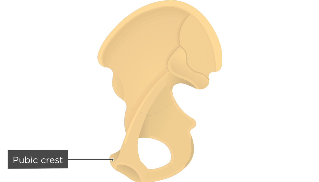
- Pubic crest (Crista pubica) is a short, superiomedial ridge that runs extends horizontally from the symphysis to the pubic tubercle. It is an attachment point for the abdominal muscles.
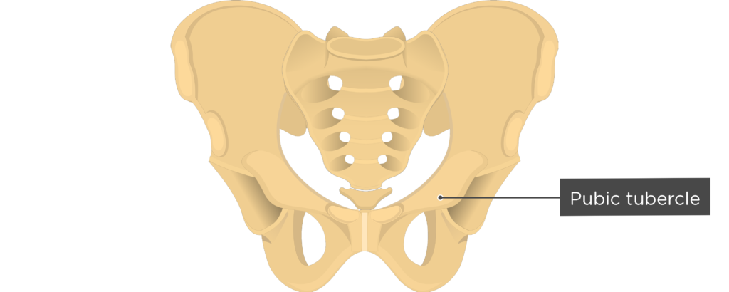
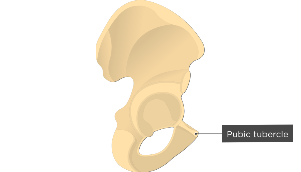
- Pubic tubercle (Tuberculum pubicum) is a projection from the lateral end of the pubic crest and serves as an attachment point for the inguinal ligament.


Ischium Bone
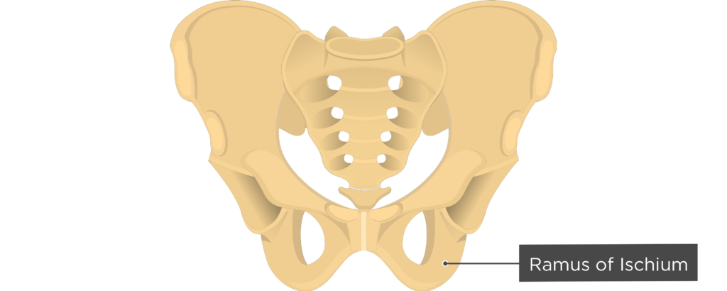
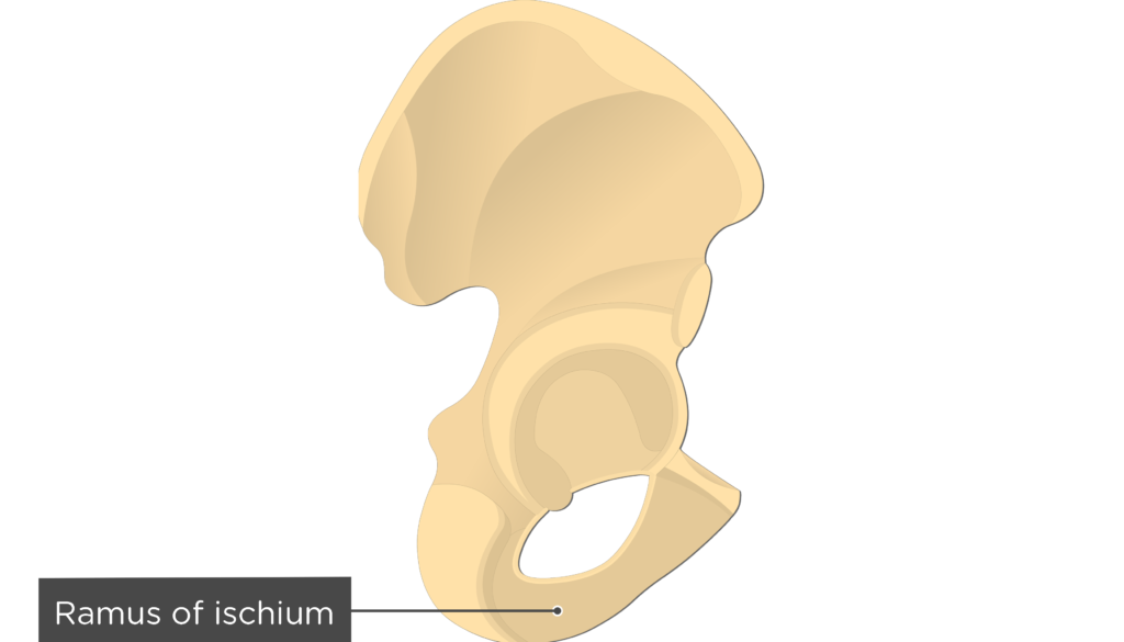
![]() Ramus of Ischium (Ramus ossis ischii) is a thin, flat, anterior extension of the ischium that joins with the inferior ramus of the pubic bone. The adductor magnus muscle attaches along its surface.
Ramus of Ischium (Ramus ossis ischii) is a thin, flat, anterior extension of the ischium that joins with the inferior ramus of the pubic bone. The adductor magnus muscle attaches along its surface.
Interdivisional Bone Markings
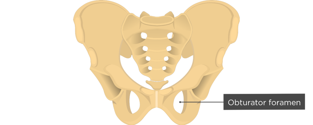
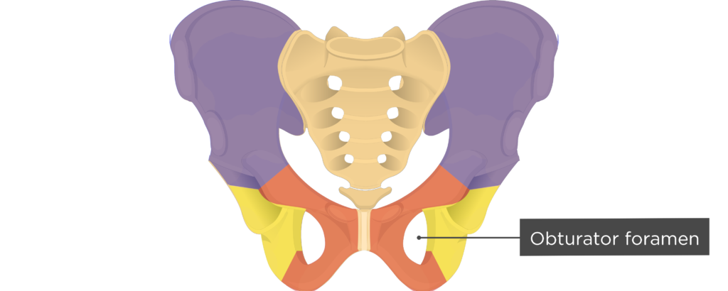
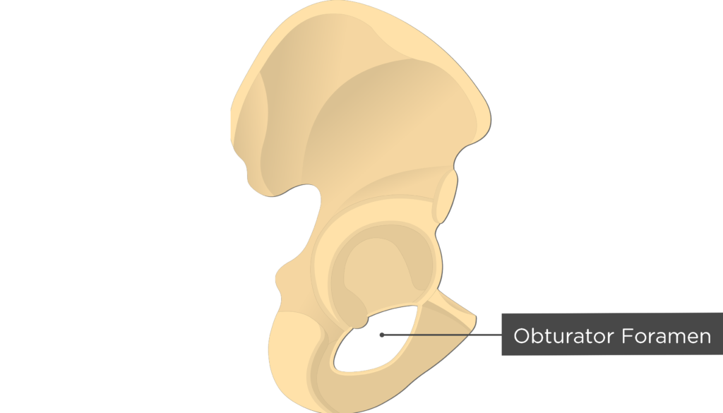
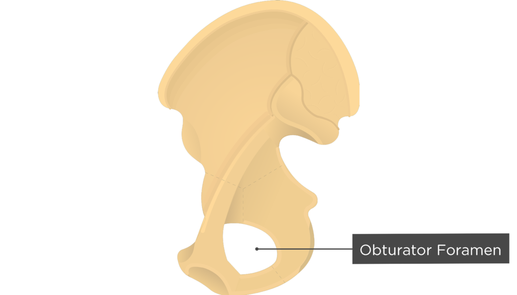
- Obturator foramen (Foramen obturatum; foramen obturatorium) is a large opening enclosed by the pubic and ischial rami. Most of the foramen is covered by a ligamentous membrane (obturator mem-brane), which helps decrease the weight of the os coax. The obturator vessels and nerves pass through a large canal located in the upper portion of the membrane.
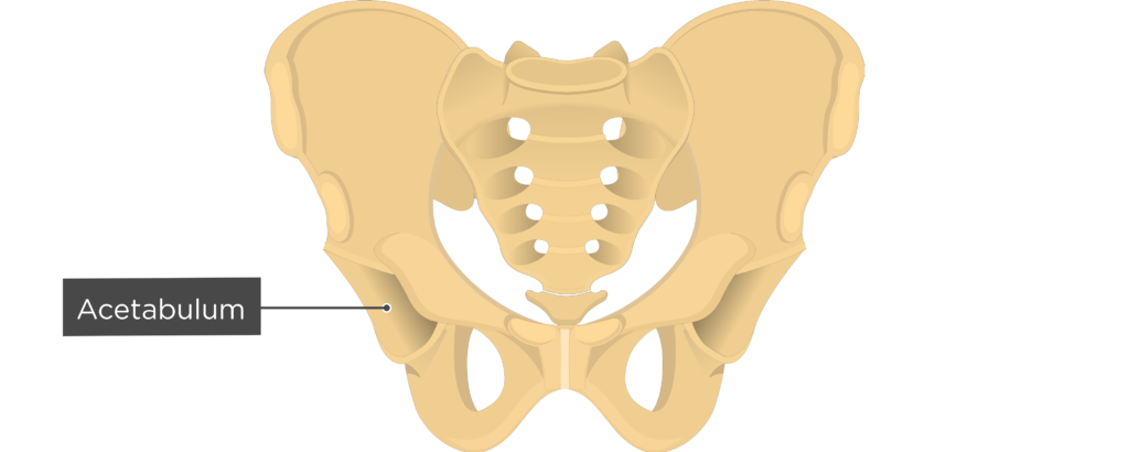
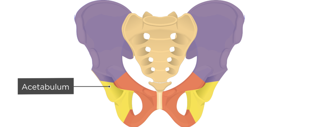
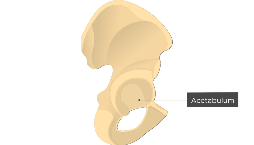
- Acetabulum (Acetabulum) is a large, rounded depression on the external (lateral) surface of the os coxa. It is formed by portions of the ilium, ischium, and pubic bones and accepts the head of the femur to form the hip joint.

Lateral or External Markings
Ilium Bone
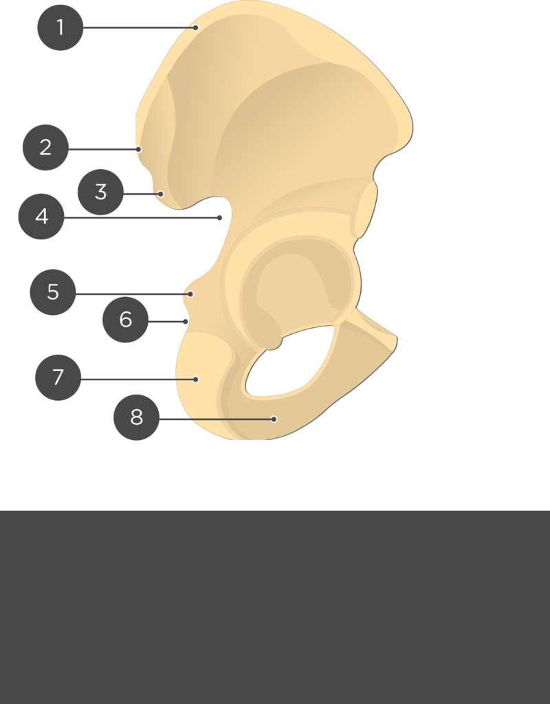
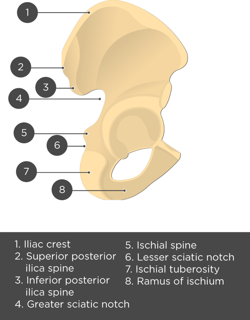
- Iliac Crest or Crest of Ilium (Crista iliaca) is the curved upper ridge of the ilium. It is an attachment point for the latissimus dorsi, quadratus lumborum, erector spine, iliacus, tensor fasciae latae, and abdominal muscles.
- Iliac Tubercle or Tubercle of the iliac Crest (Tuberculum iliac) is a prominent bulge on the outer lip of the iliac crest, near its midpoint. This area is an attachment site for the iliotibial tract or band.
- Anterior Superior Iliac Spine or ASIS (Spina iliaca anterior superior) is a projection from the anterior portion of the iliac crest. The sartorius muscle and inguinal ligament attach to this elevation.
- Anterior Inferior Iliac Spine or AIIS (Spina iliaca anterior inferior) is a projection below the anterior superior iliac spine that serves as an attachment point for the rectus femoris muscle.
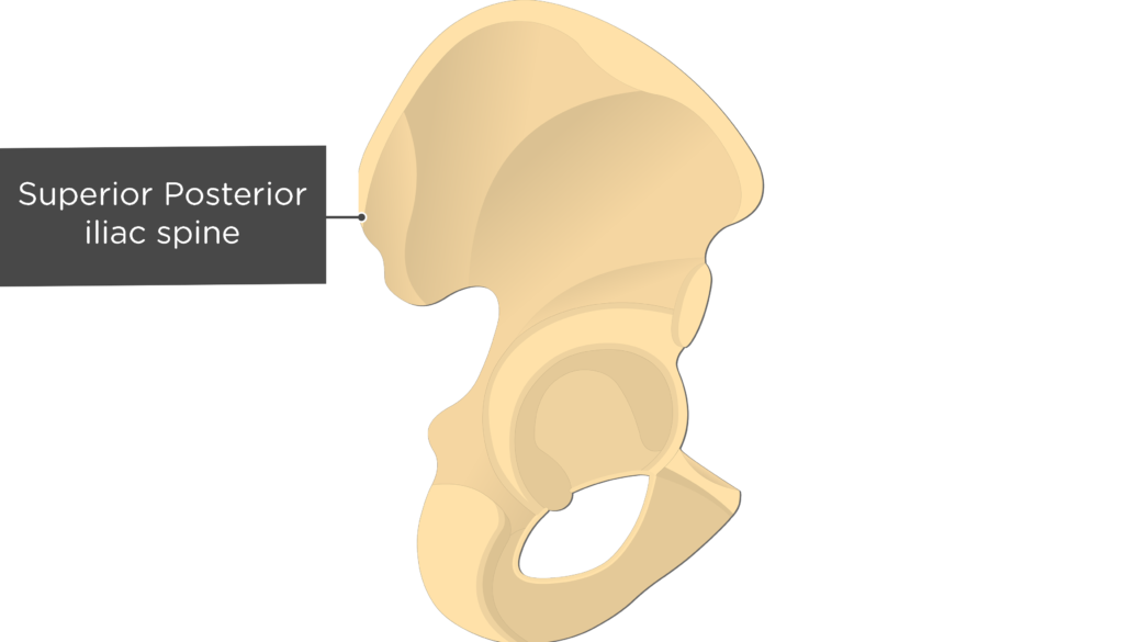
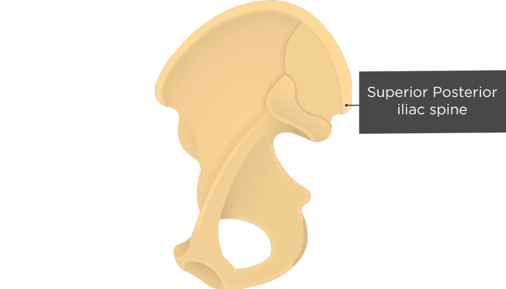
- Posterior Superior Iliac Spine (Spina iliaca posterior superior) is a projection from the posterior of the crest. It is an attachment point for the posterior sacroiliac ligament and multifidus muscle.
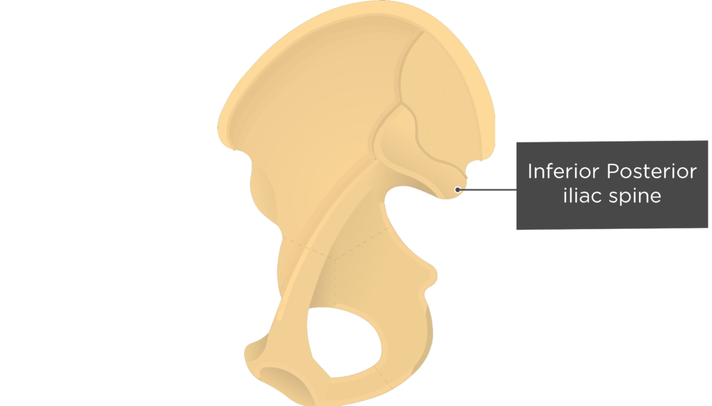
- Posterior Inferior Iliac Spine (Spina iliaca posterior inferior) is a curvature found inferior to the posterior superior iliac spine. The two spines are separated by a small notch. A ligament that binds the ilium to the sacrum attaches here.
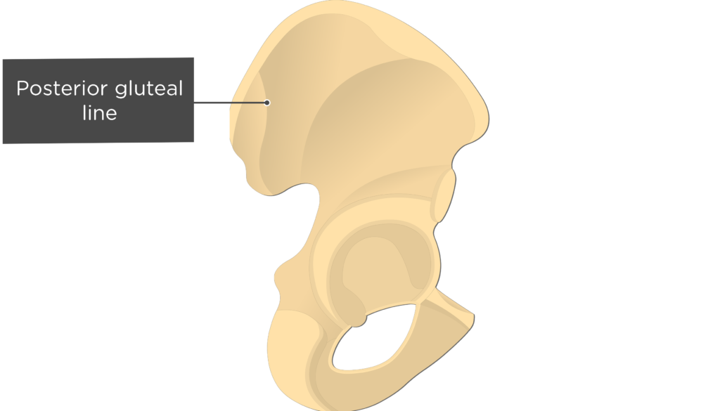
- Posterior gluteal line (Linea glutea poste-rior) is a short, ridge on the external surface of the ilium. It runs vertically between the iliac crest and greater sciatic notch, just anterior to the posterior spines. The line is the anterior attachment point for the gluteus maximus muscle and posterior attachment point for the gluteus medius muscle.
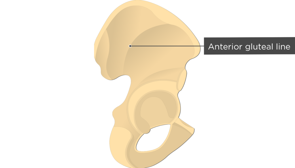
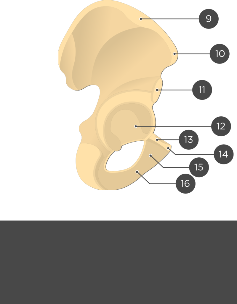
- Anterior gluteal line (Linea glutea ante-rior) is a long, posteriorly curved ridge that spans the midportion of the external ilium. The ridge serves as the anterior attachment point for the gluteus medius muscle and superior attachment point for gluteus minimus muscle.
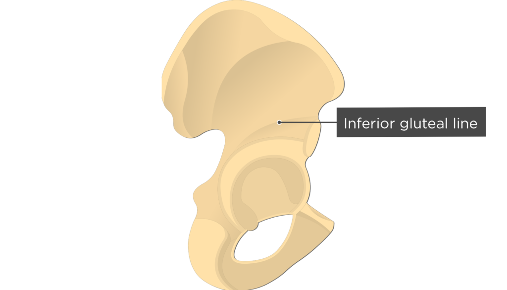
- Inferior gluteal line (Linea glutea inferior) is a slight ridge that runs diagonally across the external surface of the ilium from the anterior inferior iliac spine to the greater sciatic notch. It is the inferior attachment point for the gluteus medius muscle and superior attachment point for the gluteus minimus muscle.


Pubic Bone (Pubis)
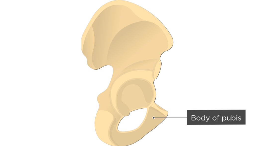
- Body of pubic bone (Corpus ossis pubis) is the flatten, medial end of the pubis that lies adjacent to the symphysis.
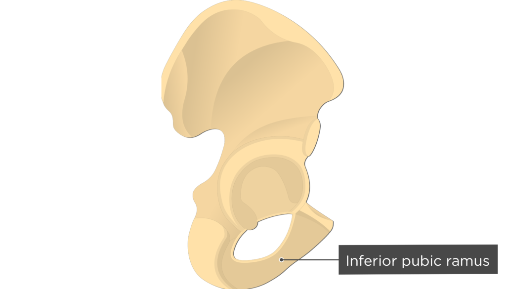
- Inferior pubic ramus (Ramus inferior ossis pubis) is a thin, flat extension from the medial pubis that joins the ischial ramus. The adductor brevis, adductor magnus, and gracilis muscles attach along this surface.
- Superior pubic ramus (Ramus superior ossis pubis) is a band of bone that runs along the superior aspect of the pubis. It is an attachment point for the pectineus muscle.
- Pubic tubercle (Tuberculum pubicum) is a projection from the lateral end of the pubic crest that serves as an attachment point for the inguinal ligament.
Ischium Bone
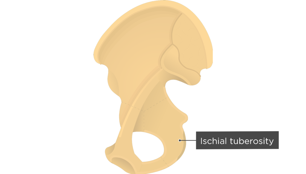
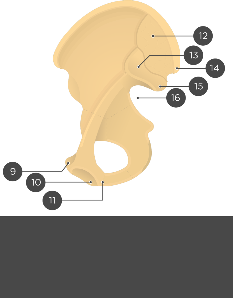
- Ischial tuberosity (Tuber ischiadicum) is a roughened, curved eminence located at the junction of the posterior and inferior borders of the ischium. It supports the weight of the body when sitting and serves as an attachment point for the sacrotuberous ligament and the hamstring muscles, quadratus femoris muscle, and inferior gemellus muscle.
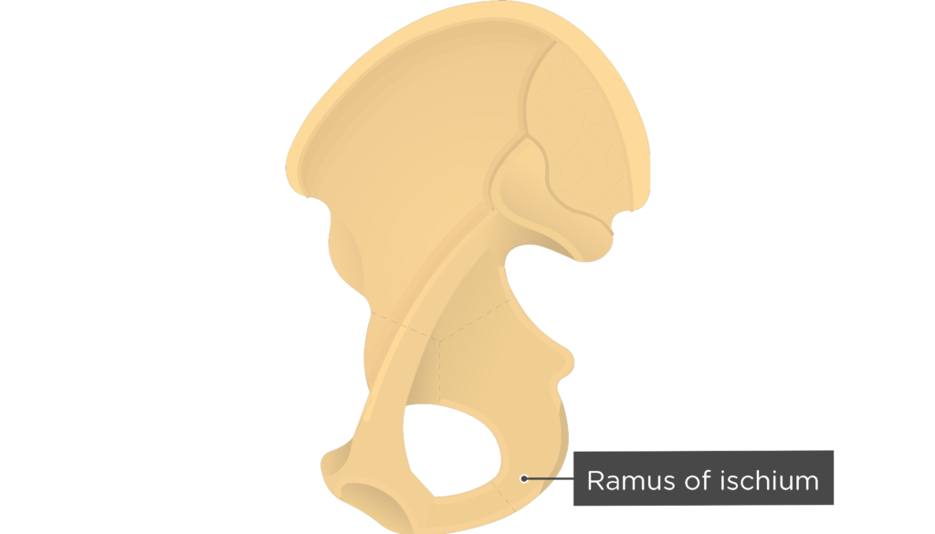
- (Inferior) Ramus of Ischium (Ramus ossis ischii) is an anterior extension from the ischial tuberosity that joins the inferior ramus of the pubic bone. The adductor magnus and obturator externus muscles attaches to this portion of the ischium.
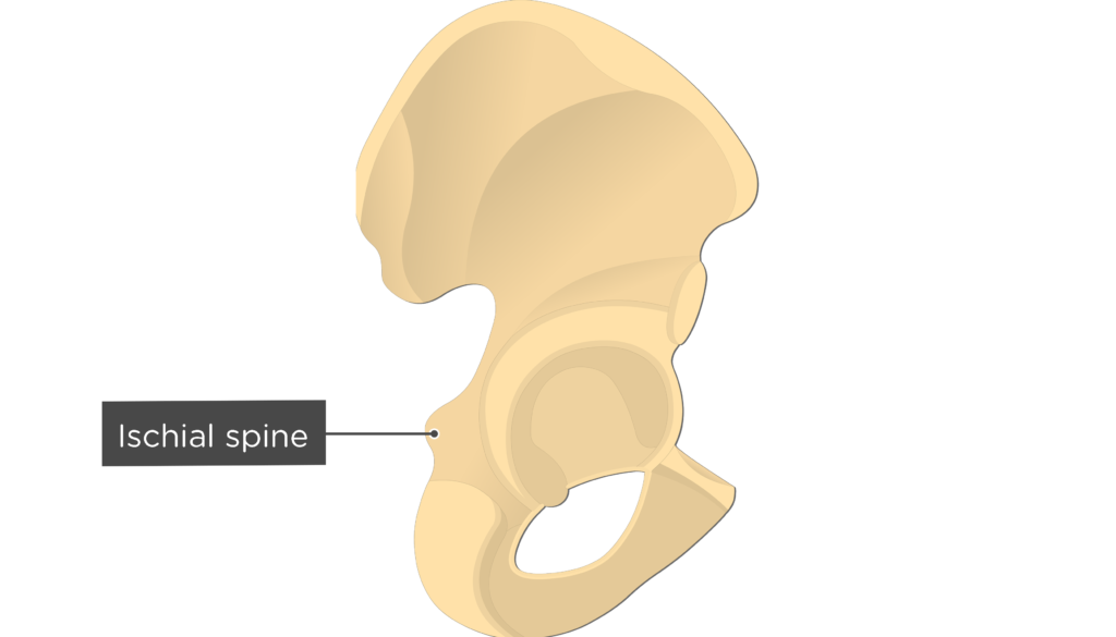
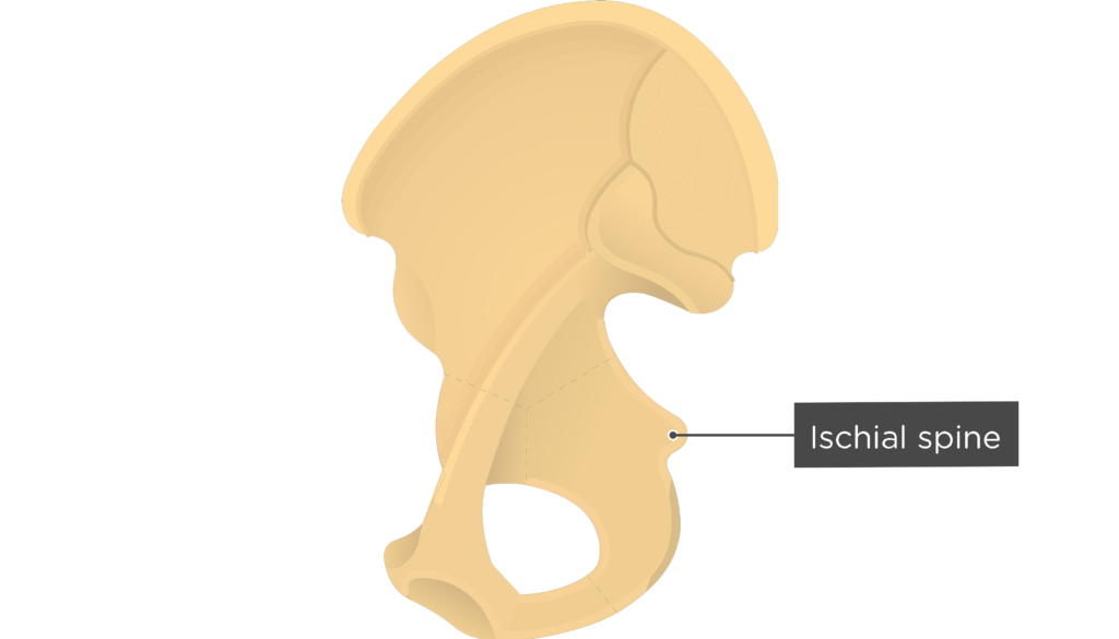
- Ischial Spine (Spina ischiadica) is a sharp projection from the posterior margin of the ischium. It is an attachment point for the sacrospinous ligament.
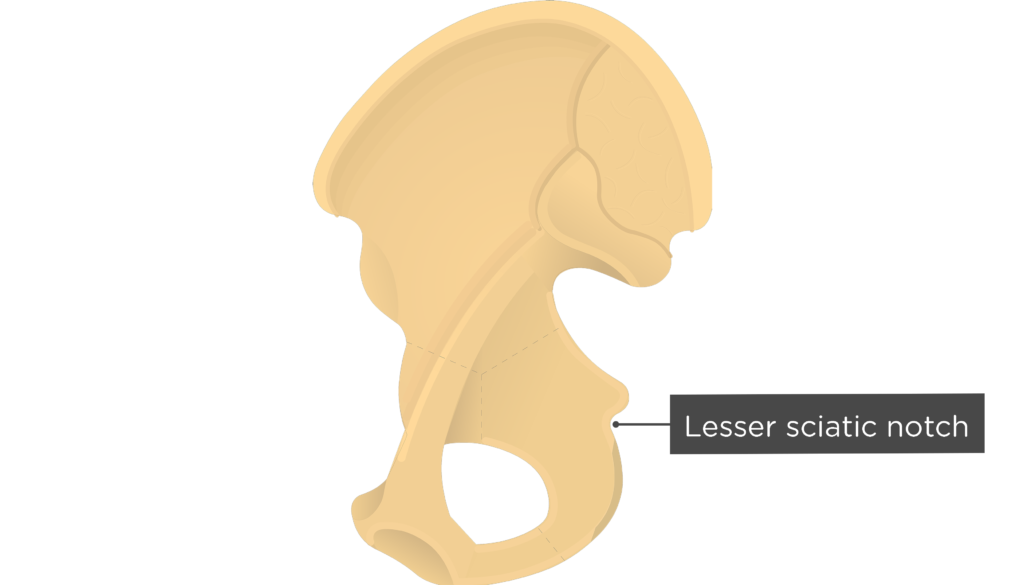
- Lesser Sciatic Notch (Incisura ischiadica minor) is a small indentation located inferior to the spine. The sacrotuberous and sacro-spinous ligaments transform the notch into the lesser sciatic foramen. This opening is a passageway for the obturator internus tendon and nerve, internal pudendal vessels, and pudendal nerve.
Interdivisional Bone Markings:
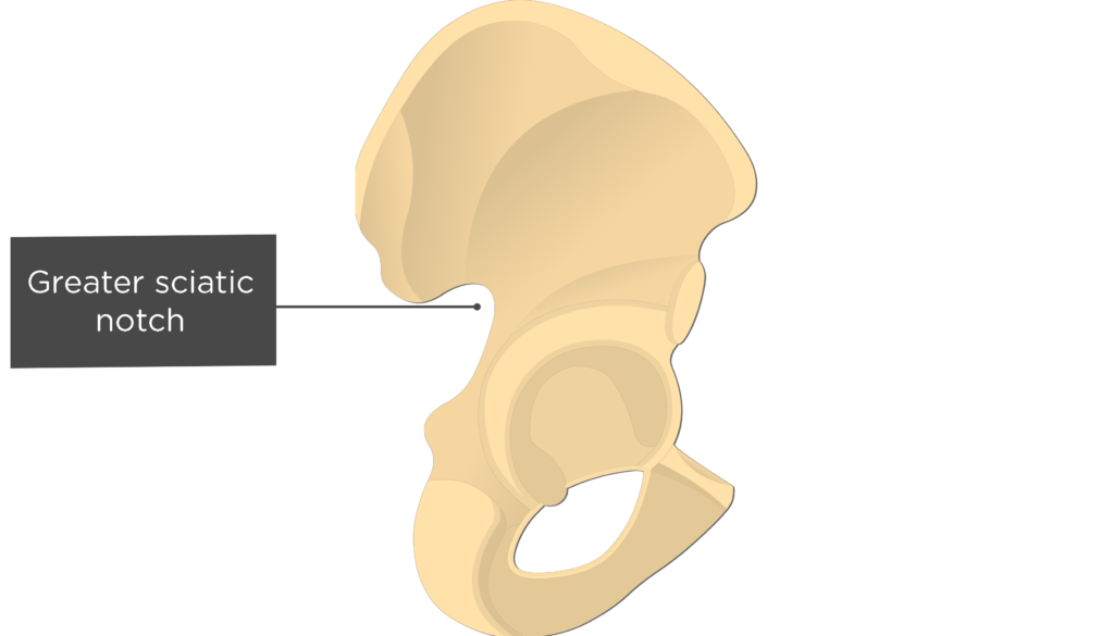
- Greater Sciatic Notch (Incisura ischiadica major) is a large indentation located below the posterior inferior iliac spine. The sacrotuberous and sacrospinous ligaments convert the notch into the greater sciatic foramen, which allows the passage of the piriformis muscle, 7 nerves (including the sciatic nerve), and 3 sets of blood vessels.
- Obturator Foramen (Foramen obturatum; foramen obturatorium) is a large anterior opening enclosed by the pubic and ischial rami. The foramen, which is mostly covered by the ligamentous obturator membrane, helps decrease the weight of the os coxae. The obturator vessels and nerves pass through a large canal located in the upper portion of the membrane.
- Acetabulum (Acetabulum) is a large, rounded depression on the external surface of the os coxa. The acetablum is formed by portions of the ilium, ischium, and pubic bones and accepts the head of the femur to form the hip joint.


Medial (Internal) Markings
Ilium Bone
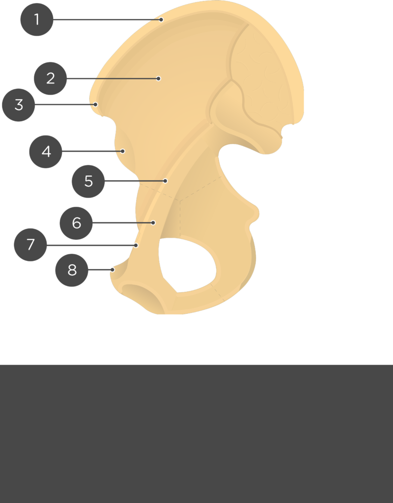
- Iliac Crest or Crest of Ilium (Crista iliaca) is the curved upper ridge of the ilium. It is an attachment point for the latissimus dorsi, quadratus lumborum, erector spine, iliacus, tensor fasciae latae, and abdominal muscles.
- Anterior Superior Iliac Spine or ASIS (Spina iliaca anterior superior) is a projection from the anterior portion of the iliac crest. The sartorius muscle and inguinal ligament attach to this elevation.
- Anterior Inferior Iliac Spine or AIIS (Spina iliaca anterior inferior) is a projection below the anterior superior iliac spine that serves as an attachment point for the rectus femoris muscle.
- Posterior Superior Iliac Spine (Spina iliaca posterior superior) is a projection from the posterior of the crest. It is an attachment point for the posterior sacroiliac ligament and multifidus muscle.
- Posterior Inferior Iliac Spine (Spina iliaca posterior inferior) is a curvature found inferior to the posterior superior iliac spine. The two spines are separated by a small notch. A ligament that binds the ilium to the sacrum attaches here.
- Iliac Fossa (Fossa iliaca) is a broad depression located along the anteromedial surface, inferior to the iliac crest. It is an attachment point for the iliacus muscle.
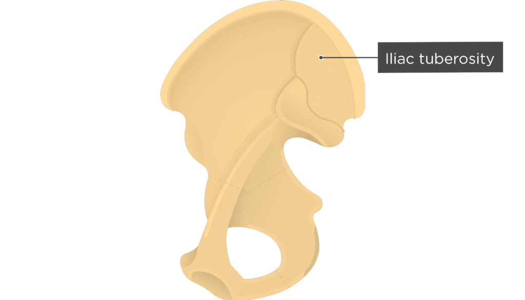
- Iliac tuberosity (Tuberositas iliaca) is a roughened area located inferior to the crest and posterior to the iliac fossa. The posterior sacroiliac ligaments and the sacrospinalis and multifidus muscles attach here.
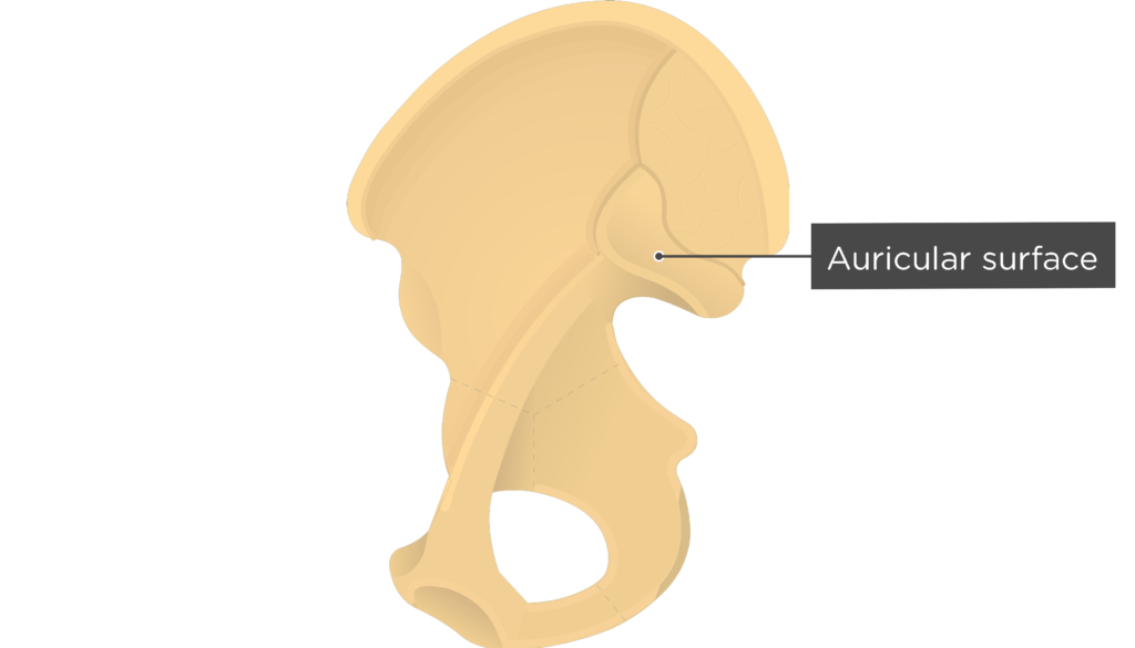
- Auricular surface (Facies auricular ossis ilii) is a L-shaped or ear-shaped roughened surface situated inferior to the tuberosity. This area articulates with auricular surface of sacrum to form the sacroiliac joint.
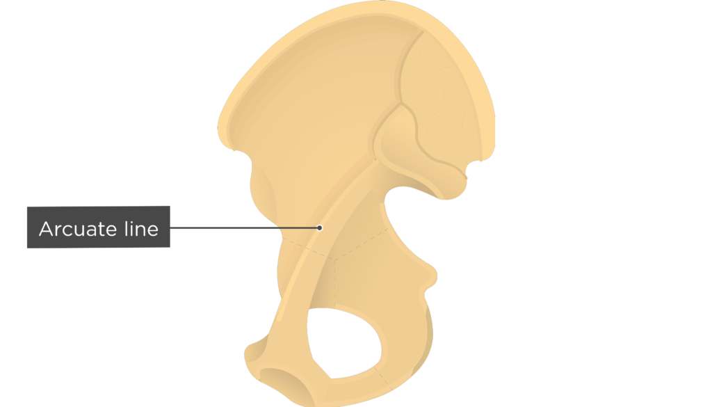
- Arcuate line (Linea arcuata) is a curved ridge that forms the inferior boundary of the iliac fossa. It also delinates the boundary between the body and the wing (large expanded portion; ala) of the ilium.
Pubic Bone (Pubis)
- Symphysis pubis or pubic symphysis (Symphysis pubica; symphysis pubis) is the cartilaginous joint between the two pubic bones.
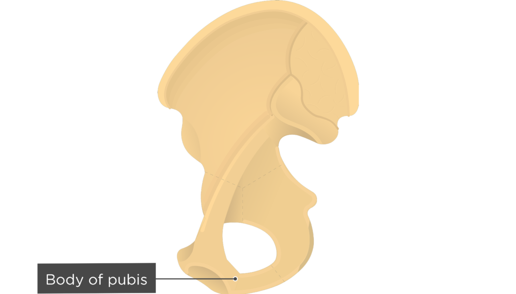
- Body of pubis (Corpus ossis pubis) is the flatten, medial end of the pubis that lies adjacent to the symphysis.
- Inferior pubic ramus (Ramus inferior ossis pubis) is a thin, flat extension from the medial pubis that joins the ischial ramus. The adductor brevis, adductor magnus, and gracilis muscles attach along this surface.
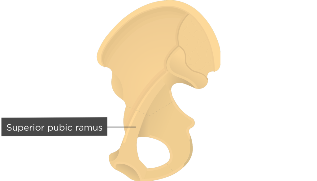
- Superior pubic ramus (Ramus superior ossis pubis) is a band of bone that runs along the superior aspect of the pubis. It is an attachment point for the pectineus muscle.
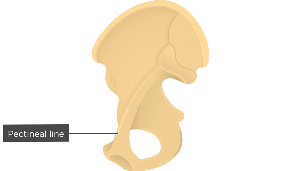
- Pectineal line (Linea pectinea; pecten ossis pubis) is a sharp ridge that runs along the superior margin of the superior pubic ramus. This area, which is also called the pecten pubis, is an the attachment point for the pectineus muscle.
- Pubic crest (Crista pubica) is a short, superiomedial ridge that runs extends horizontally from the symphysis to the pubic tubercle. It is an attachment point for the abdominal muscles.
- Pubic tubercle (Tuberculum pubicum) is a projection from the lateral end of the pubic crest that serves as an attachment point for the inguinal ligament.


Ischium Bone
- Ischial tuberosity (Tuber ischiadicum) is a roughened, curved eminence located at the junction of the posterior and inferior borders of the ischium. It supports the weight of the body when sitting and serves as an attachment point for the sacrotuberous ligament and the hamstring muscles, quadratus femoris muscle, and inferior gemellus muscle.
- (Inferior) Ramus of Ischium (Ramus os-sis ischii) is an anterior extension from the ischial tuberosity that joins the inferior pubic ramus. The adductor magnus and obturator externus muscles attach to this surface.
- Ischial Spine (Spina ischiadica) is a sharp projection from the posterior margin of the ischium. It is an attachment point for the sacrospinous ligament.
- Lesser Sciatic Notch (Incisura ischiadica minor) is a indentation located below the spine. The sacrotuberous and sacrospinous ligaments transform the notch into the lesser sciatic foramen, which allows the obturator internus tendon and nerve, internal pudendal vessels, and pudendal nerve to pass.
Interdivisional Bone
- Greater Sciatic Notch (Incisura ischiadica major) is a large indentation located below the posterior inferior iliac spine. The sacrotuberous and sacrospinous ligaments transform the notch into the greater sciatic foramen, which allows the passage of the performs muscle, 7 nerves (including the sciatic nerve), and 3 sets of blood vessels.
- Obturator Foramen (Foramen obturatum; foramen obturatorium) is a large anterior opening enclosed by the pubic and ischial rami. The foramen, which is mostly covered by a ligamentous membrane, helps decrease the weight of the os coxae. The obturator vessels and nerves pass through a large canal in the upper portion of the membrane.

Test yourself
- ِAnterior view: [Show/Hide]
- Lateral view – Part 1: [Show/Hide]
- Lateral view – Part 2: [Show/Hide]
- Medial view – Part 1: [Show/Hide]
- Medial view – Part 1: [Show/Hide]


Interactive quizzes about the hip bone (os coxa)
Os Coxa Bone Quiz – Anterior Markings
Os Coxa Bone Quiz – Lateral View Markings
Os Coxa Bone Quiz – Medial View Markings
Related Articles

Tibia and Fibula Bones – Anatomy
An introduction to the tibia and fibula bones of the leg. Learn about the different markings and test yourself.
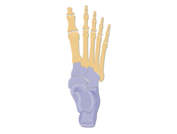
Tarsals | Tarsal Bones Anatomy
The tarsal bones are seven bones articulating with each other. They build the connection between the lower leg and the metatarsus.
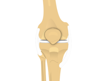
Patella Bone – Anterior and Posterior Views
The patella, also known as the kneecap, is a triangular shaped bone located anterior to a groove between the femur condyles called the patellar surface. It covers and protects the distal surface of the anterior femur and functions to displace the quadriceps tendon away from the femurotibial joint surface.
