Extensor Carpi Radialis Brevis Muscle
Author: Dimitrios Mytilinaios MD, PhD

Last update:

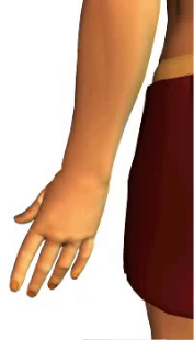
The extensor carpi radialis brevis is a muscle found on the lateral side of the forearm. Anatomically, it belongs to a vast group of superficial extensors of the forearm, together with anconeus, brachioradialis, extensor carpi radialis longus, extensor digitorum, extensor digiti minimi and extensor carpi ulnaris.

The function of this muscle is to extend the hand. In the following text we will describe its origin, insertion, as well as innervation and functions.

Extensor carpi radialis brevis muscle
OriginLateral epicondyle of the humerus
InsertionBase of the third metacarpal bone
InnervationRadial nerve
FunctionsExtension of the hand;
Abduction of the hand (radial deviation)

image descriptionExtensor Carpi Radialis Brevis Muscle
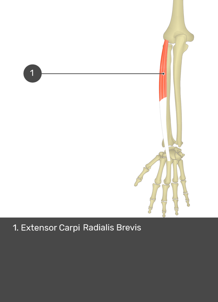
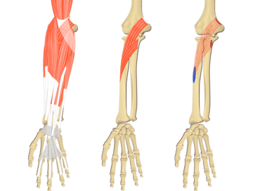
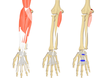
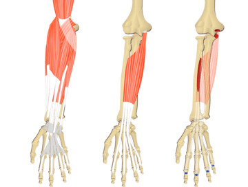
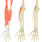
A posterior view of the forearm showing the bony elements and isolated Extensor Carpi Radialis Brevis muscle.
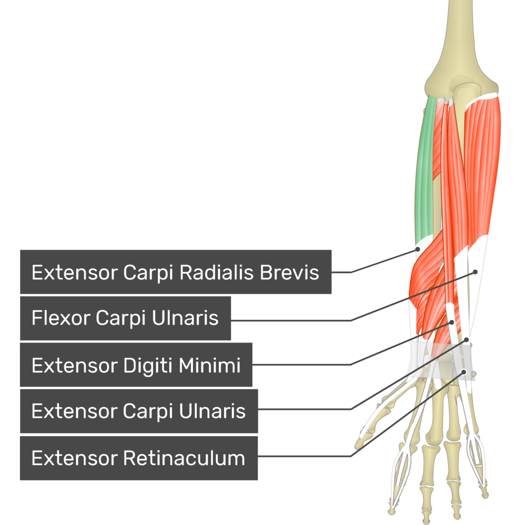
A posterior view of the forearm showing the bony elements and the deeper muscles. The visible, labelled muscles are as follows: Triceps Brachii, Brachioradialis, Extensor Carpi Radialis Longus, Anconeus, Extensor Digitorum, Extensor Carpi Radialis Brevis (highlighted in green), Flexor Carpi Ulnaris, Extensor Digiti Minimi, Extensor Carpi Ulnaris and Extensor Retinaculum.
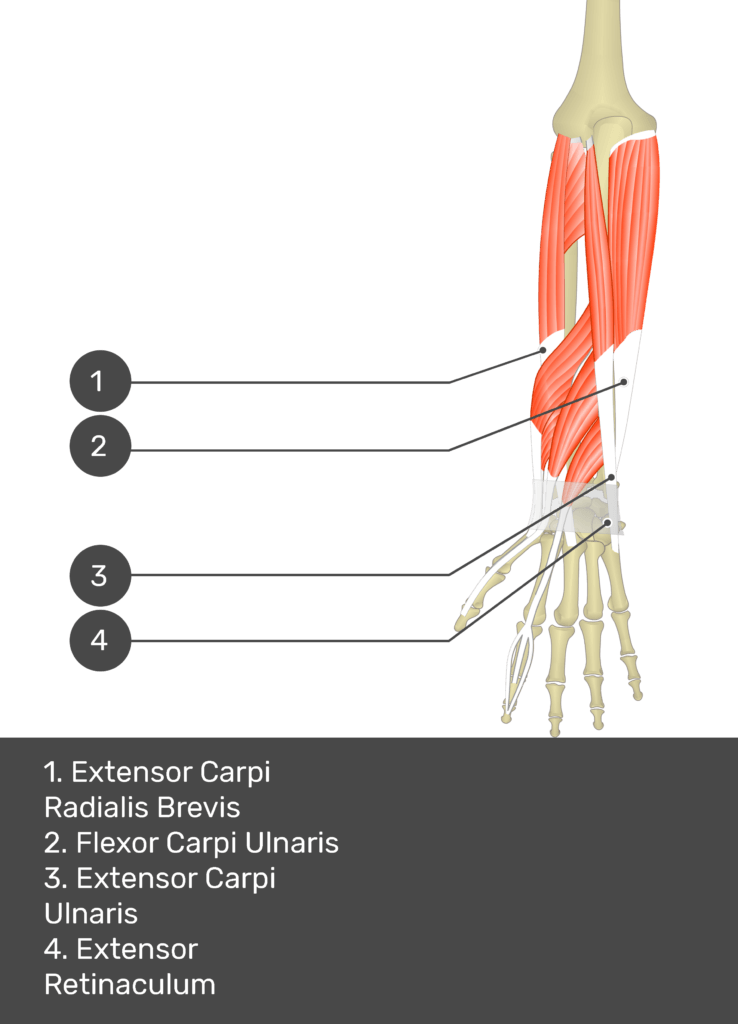
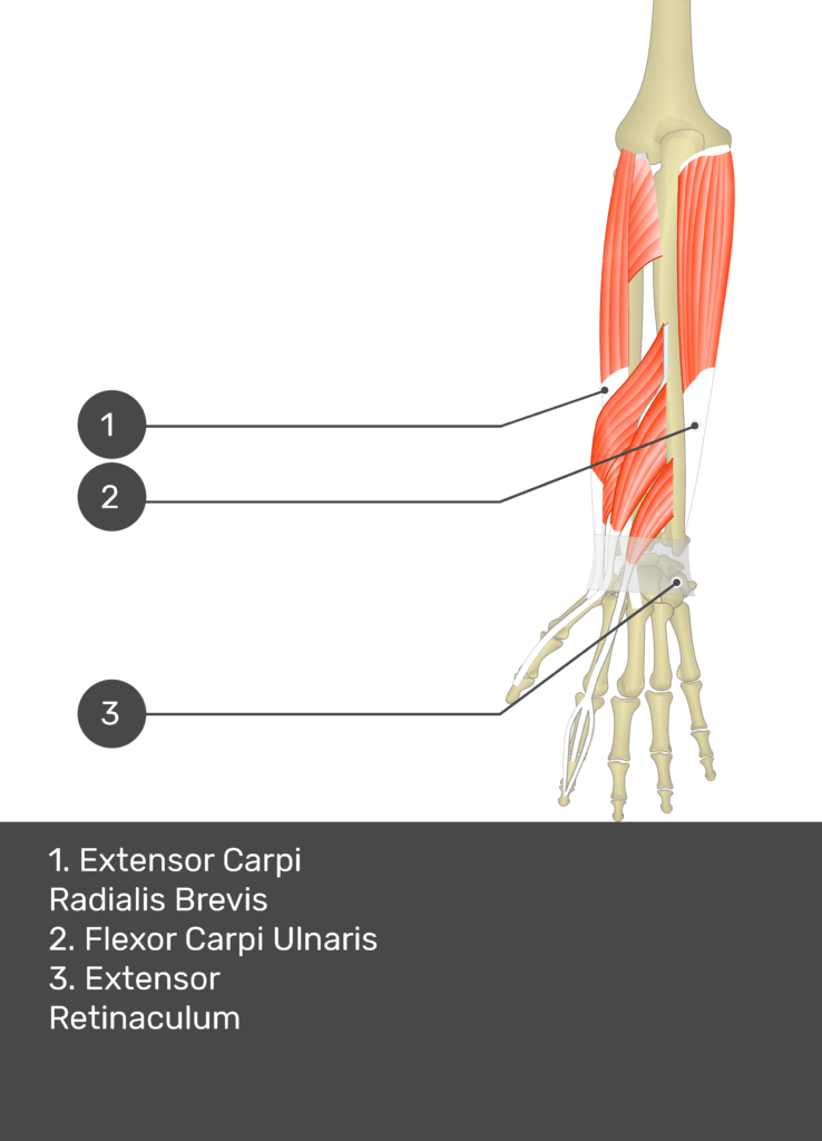
A posterior view of the forearm showing the bony elements and the deeper muscles. The visible, labelled muscles are as follows: Extensor Carpi Radialis Brevis (highlighted in green), Flexor Carpi Ulnaris, Extensor Digiti Minimi, Extensor Carpi Ulnaris and Extensor Retinaculum.
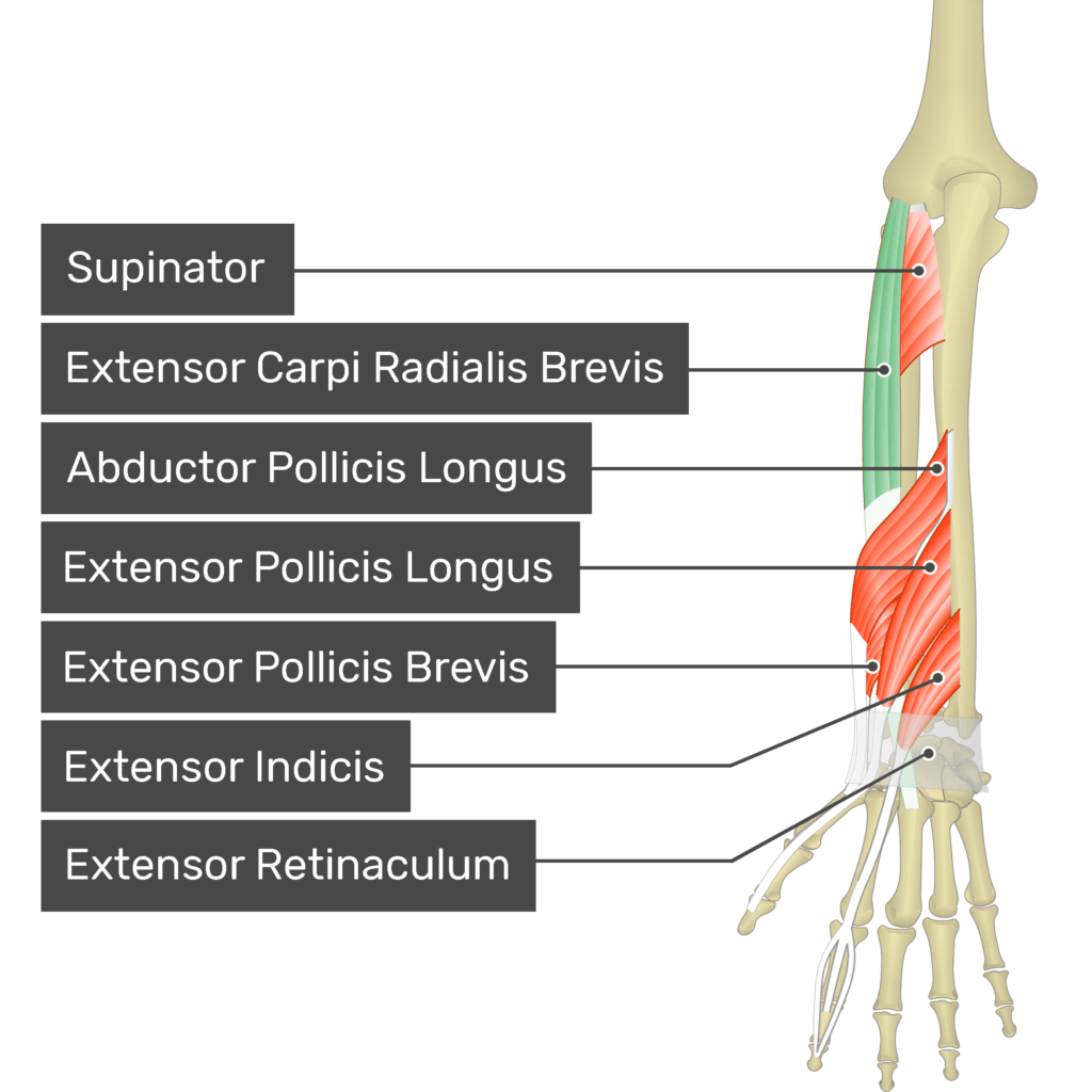
A posterior view of the forearm showing the bony elements and the deeper muscles. The visible, labelled muscles are as follows: Supinator, Extensor Carpi Radialis Brevis (highlighted in green), Abductor Pollicis Longus, Extensor Pollicis Longus, Extensor Pollicis Brevis, Extensor Indicis and Extensor Retinaculum.
1
2
3
4

Origin and insertion

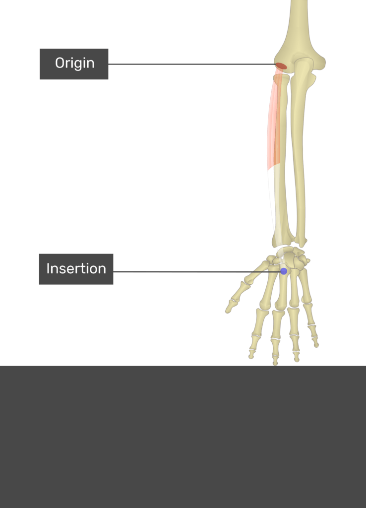
The extensor carpi radialis brevis originates from the lateral epicondyle of the humerus. Its fusiform muscle belly then extends downwards and around the midpoint of the forearm it gives rise to a thin tendon. The tendon crosses the posterior side of the wrist joint and enters the hand, where it inserts to the base of the 3rd metacarpal bone.

image descriptionAttachments of the extensor carpi radialis brevis muscle

A diagram depicting the origin and insertion points of the extensor carpi radialis brevis muscle.
1

Learning the muscles of the forearm is tough! Save time and energy with these upper limb muscle anatomy reference charts.

Functions

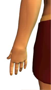
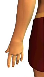
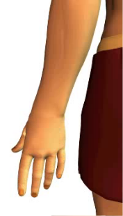
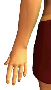
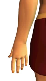
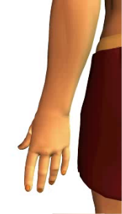
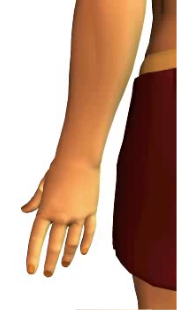
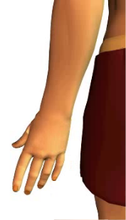
The main function of the extensor carpi radialis is to act on the wrist joint. When it contracts together with the extensor carpi ulnaris, it produces the image descriptionextension of the hand. This is because the extensor carpi ulnaris is found on the opposite side of the forearm (medial), so its pull balances out the pull of the extensor carpi radialis brevis.

Slide 1 of the animation of wrist extension
Slide 2 of the animation of wrist extension
Slide 3 of the animation of wrist extension
Slide 4 of the animation of wrist extension
Slide 5 of the animation of wrist extension
Extension of the hand

On the other hand, when the extensor carpi radialis contracts together with other muscles found on the lateral side of the forearm, namely the extensor carpi radialis longus and flexor carpi radialis, it pulls the hand medially producing the image descriptionabduction of the hand, also called the radial deviation of the hand.

Slide 1 of the wrist abduction animation
Slide 2 of the wrist abduction animation
Slide 3 of the wrist abduction animation
Slide 4 of the wrist abduction animation
Slide 5 of the wrist abduction animation
Abduction (radial deviation) of the hand

Innervation

The extensor carpi radialis brevis is innervated by the radial nerve (C6, C7).

Posterior forearm muscles

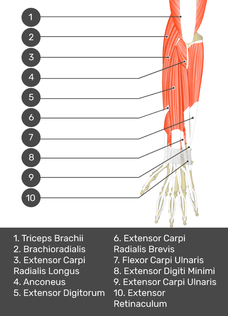
A test yourself image of the dorsal view of the forearm showing the bony elements and the deeper muscles. The visible muscles of the forearm are numbered 1-10 and the answers in the box below are concealed.
A test yourself image of the dorsal view of the forearm showing the bony elements and the deeper muscles. The visible muscles of the forearm are numbered 1-10. The answers in the box below are as follows 1. Triceps Brachii 2. Brachioradialis 3. Extensor Carpi Radialis Longus 4. Anconeus 5. Extensor Digitorum 6. Extensor Carpi Radialis Brevis 7. Flexor Carpi Ulnaris 8. Extensor Digiti Minimi 9. Extensor Carpi Ulnaris 10. Extensor Retinaculum.
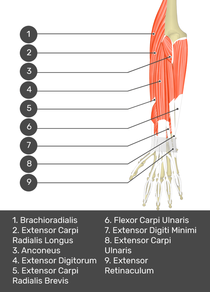
A test yourself image of the dorsal view of the forearm showing the bony elements and the deeper muscles. The visible muscles of the forearm are numbered 1-9. The answers in the box below are as follows 1. Brachioradialis 2. Extensor Carpi Radialis Longus 3. Anconeus 4. Extensor Digitorum 5. Extensor Carpi Radialis Brevis 6. Flexor Carpi Ulnaris 7. Extensor Digiti Minimi 8. Extensor Carpi Ulnaris 9. Extensor Retinaculum.
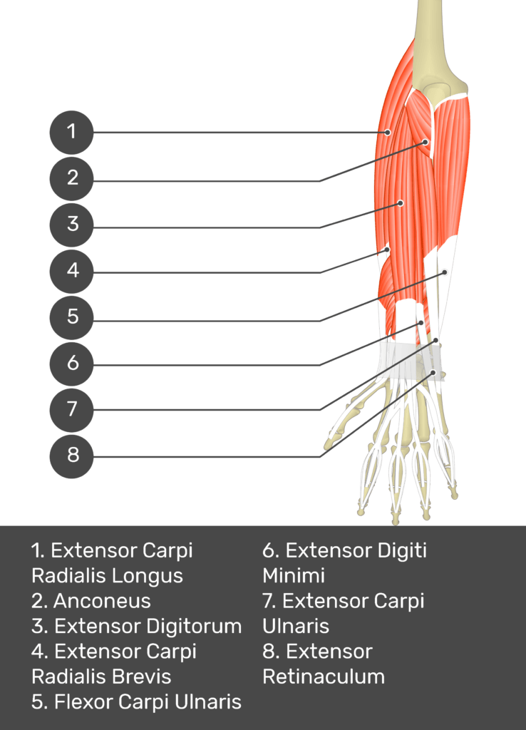
A test yourself image of the dorsal view of the forearm showing the bony elements and the deeper muscles. The visible muscles of the forearm are numbered 1-8. The answers in the box below are as follows 1. Extensor Carpi Radialis Longus 2. Anconeus 3. Extensor Digitorum 4. Extensor Carpi Radialis Brevis 5. Flexor Carpi Ulnaris 6. Extensor Digiti Minimi 7. Extensor Carpi Ulnaris 8. Extensor Retinaculum.
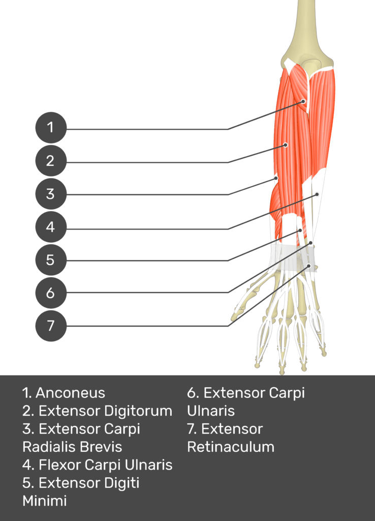
A test yourself image of the dorsal view of the forearm showing the bony elements and the deeper muscles. The visible muscles of the forearm are numbered 1-7. The answers in the box below are as follows 1. Anconeus 2. Extensor Digitorum 3. Extensor Carpi Radialis Brevis 4. Flexor Carpi Ulnaris 5. Extensor Digiti Minimi 6. Extensor Carpi Ulnaris 7. Extensor Retinaculum.
A test yourself image of the dorsal view of the forearm showing the bony elements and the deeper muscles. The visible muscles of the forearm are numbered 1-6. The answers in the box below are as follows 1. Extensor Digitorum 2. Extensor Carpi Radialis Brevis 3. Flexor Carpi Ulnaris 4. Extensor Digiti Minimi 5. Extensor Carpi Ulnaris 6. Extensor Retinaculum.
A test yourself image of the dorsal view of the forearm showing the bony elements and the deeper muscles. The visible muscles of the forearm are numbered 1-5. The answers in the box below are as follows 1. Extensor Carpi Radialis Brevis 2. Flexor Carpi Ulnaris 3. Extensor Digiti Minimi 4. Extensor Carpi Ulnaris 5. Extensor Retinaculum.
A test yourself image of the dorsal view of the forearm showing the bony elements and the deeper muscles. The visible muscles of the forearm are numbered 1-4. The answers in the box below are as follows 1. Extensor Carpi Radialis Brevis 2. Flexor Carpi Ulnaris 3. Extensor Carpi Ulnaris 4. Extensor Retinaculum.
A test yourself image of the dorsal view of the forearm showing the bony elements and the deeper muscles. The visible muscles of the forearm are numbered 1-3. The answers in the box below are as follows 1. Extensor Carpi Radialis Brevis 2. Flexor Carpi Ulnaris 3. Extensor Retinaculum.
A test yourself image of the dorsal view of the forearm showing the bony elements and the deeper muscles. The visible muscles of the forearm are numbered 1-7. The answers in the box below are as follows 1. Supinator 2. Extensor Carpi Radialis Brevis 3. Abductor Pollicis Longus 4. Extensor Pollicis Longus 5. Extensor Pollicis Brevis 6. Extensor Indicis 7. Extensor Retinaculum.
A test yourself image of the dorsal view of the forearm showing the bony elements and the isolated Extensor Carpi Radialis Brevis muscle numbered 1.
A test yourself image of the dorsal view of the forearm showing the bony elements and the attachments of the Extensor Carpi Radialis Brevis muscle. The origin at the lateral epicondyle of humerus is marked by a red circle and the insertion at the base of 3rd metacarpal (dorsal surface) is marked by a blue oval.
Test yourself while observing the Extensor Carpi Radialis Brevis muscle
1
2
3
4
5
6
7
8
9
10
11
12

References

  • Palastanga, N., & Soames, R. (2012). Anatomy of Human Movement (6th ed.). Edinburgh, Scotland: Churchill Livingstone.
  • Open Anatomy. (n.d.). TA2 Viewer. Retrieved April 5, 2023, from https://ta2viewer.openanatomy.org/
  • Moore, K. L. (2018). Clinically Oriented Anatomy (8th ed.). Philadelphia, PA: Wolters Kluwer.
  • Drake, R. L., Vogl, A. W., & Mitchell, A. W. M. (2015). Gray’s Anatomy for Students (3rd ed.). Edinburgh, Scotland: Churchill Livingstone.
  • Standring, S. (2021). Gray’s Anatomy (42tst ed.). Edinburgh: Elsevier Churchill Livingstone.