Flexor Carpi Radialis
Pronator Teres – Attachments, Action & Innervation
Last update:
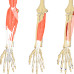
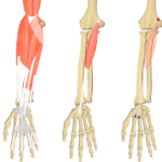
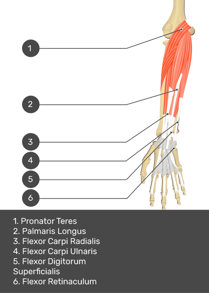
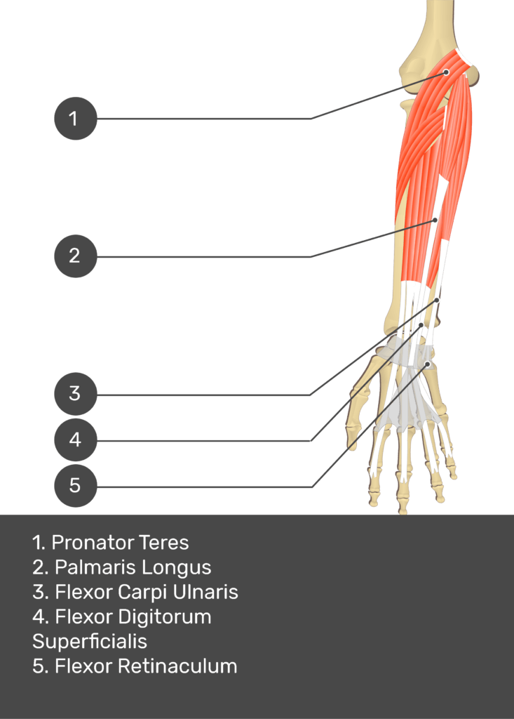
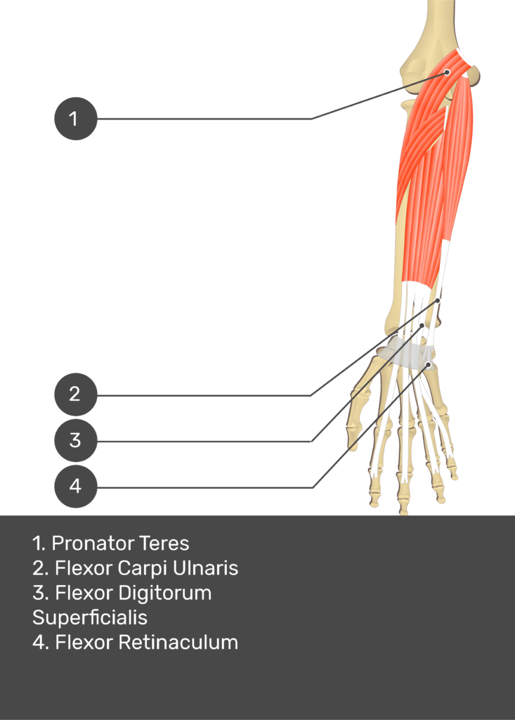
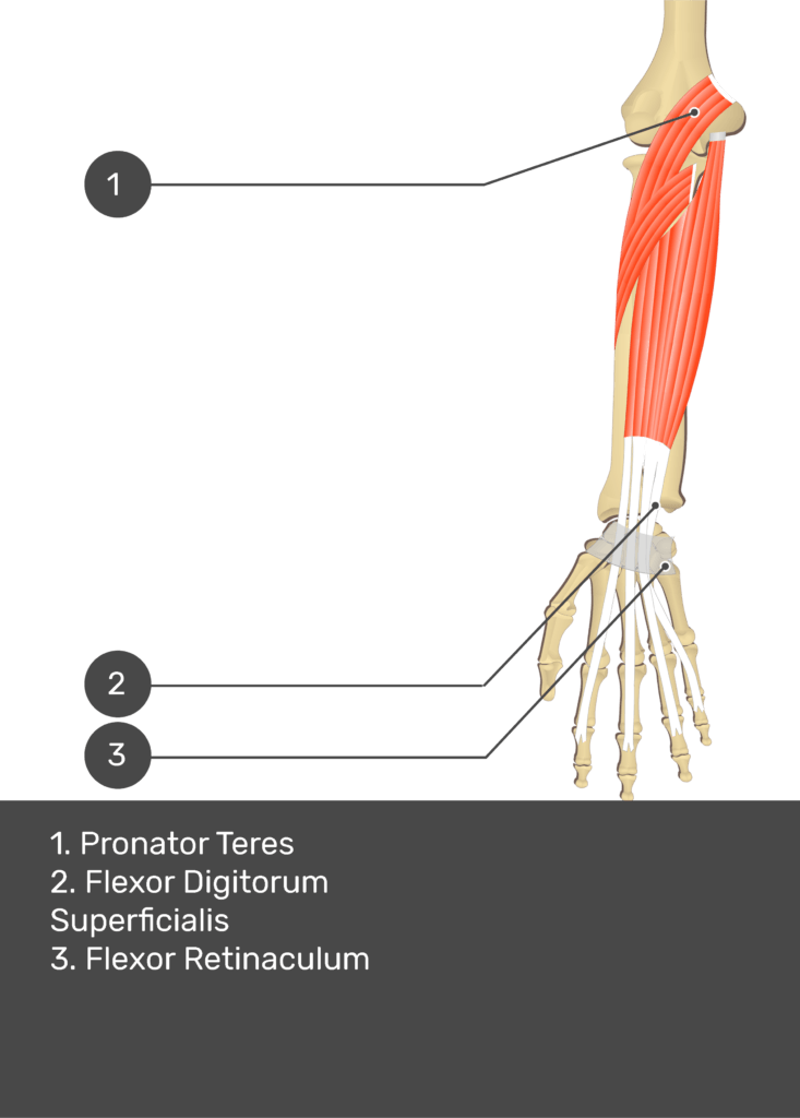
Pronator teres is one of the muscles of the superficial flexor group of the anterior forearm compartment.
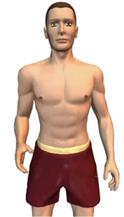
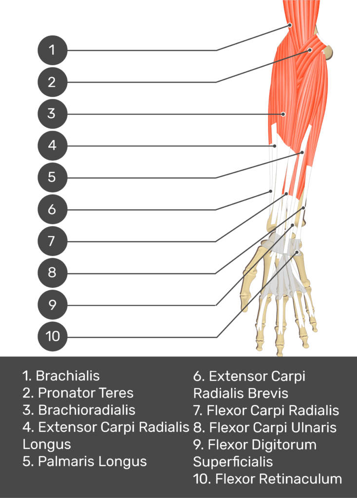
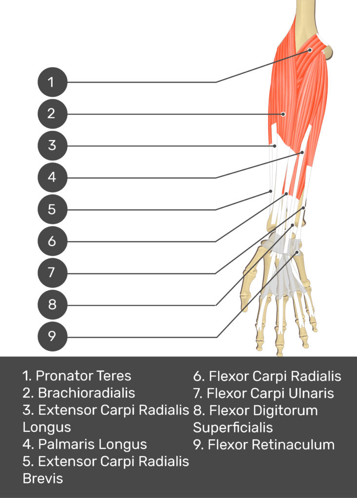
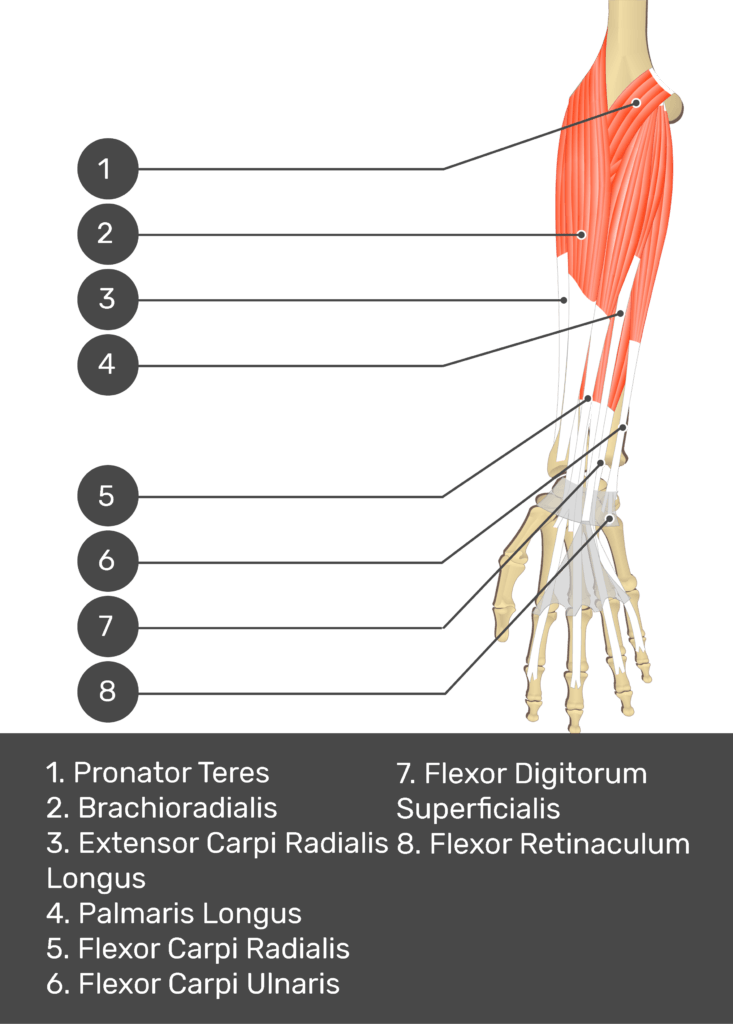
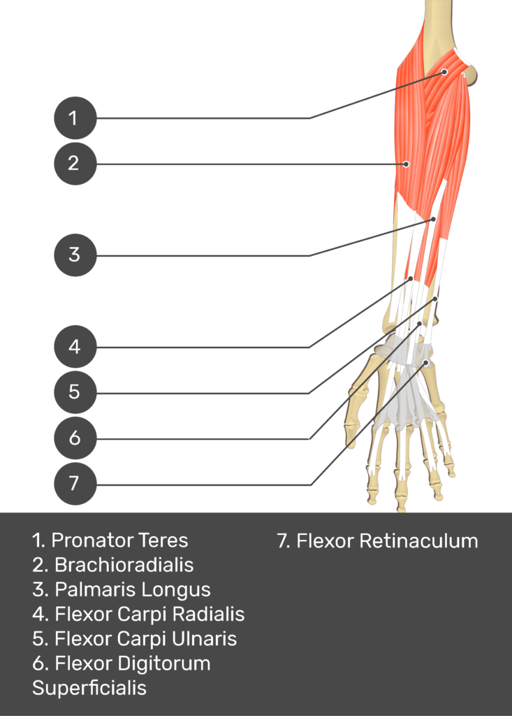
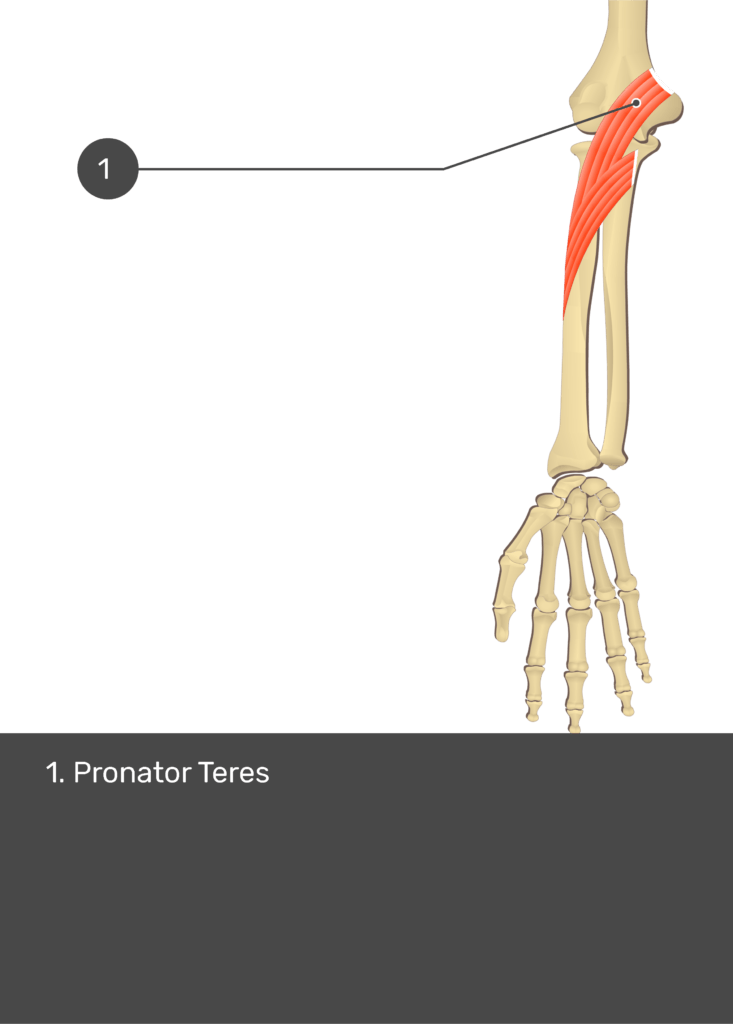
 Pronator Teres Muscle


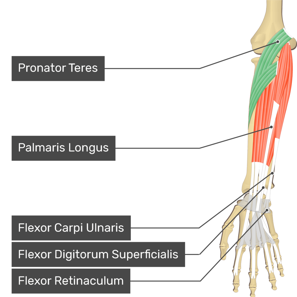

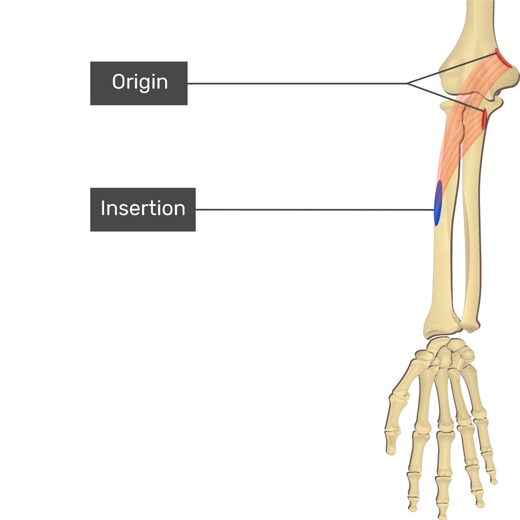
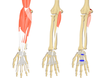
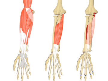
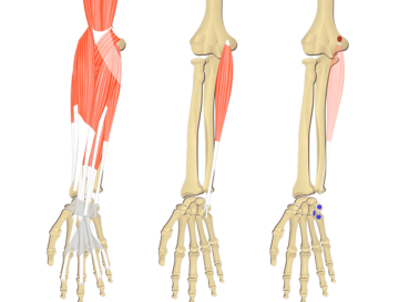
Attachments of Pronator Teres Muscle: Origin and Insertion
Origin (proximal attachments):
a. Humoral head – medial epicondyle of humerus & distal supracondylar ridge
b. Ulnar head – medial side of coronoid process of ulna.
Insertion (distal attachment):
a. Middle of lateral surface of radius.
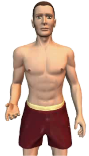
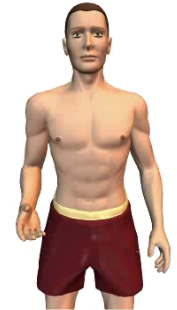
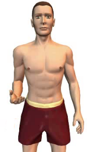
Actions of Pronator Teres Muscle
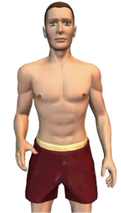
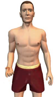
a. ![]() Pronates the forearm at the elbow.
Pronates the forearm at the elbow.
Learn the anatomy of the pronator teres muscle in half the time with this muscle anatomy reference guide.
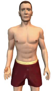
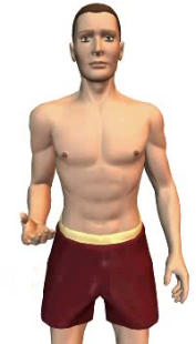
b. ![]() Flexes the forearm at the elbow.
Flexes the forearm at the elbow.
Nerve to Pronator Teres muscle and its spinal segment:
a. Median nerve (C6, C7).
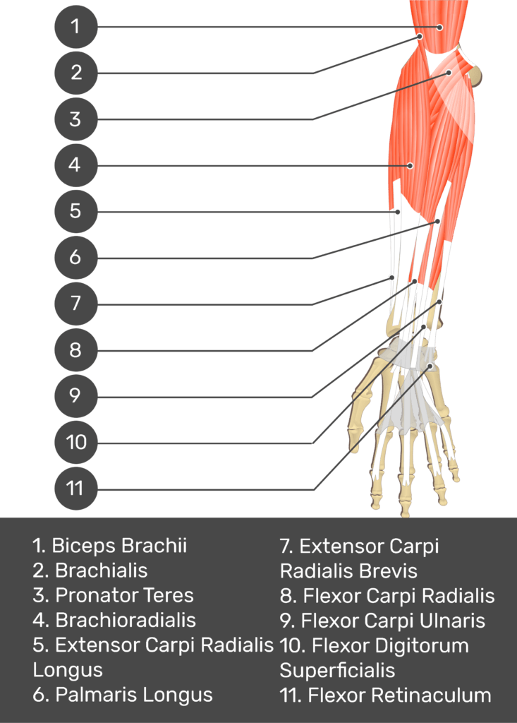
An Overview of the Anterior Forearm Muscles
Muscles That Act On The Anterior Forearm
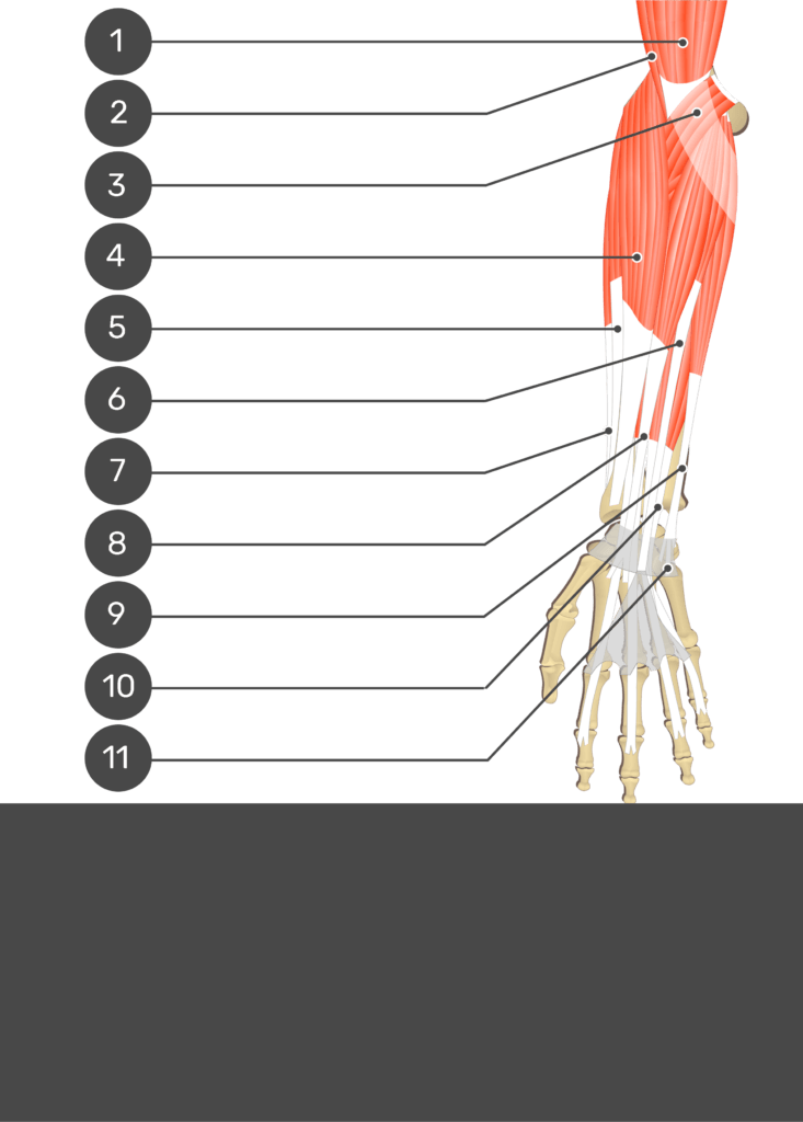

Test yourself while observing the Pronator Teres muscle.
Related Articles
Palmaris Longus Muscle
A tutorial on the position, actions, attachments and innervation of the Palmaris Longus muscle with the aid of detailed anatomical illustrations and a quiz.
Flexor Digitorum Superficialis Muscle
A tutorial on the position, actions, attachments and innervation of the Flexor Digitorum Superficialis muscle with the aid of anatomical illustrations.
Flexor Carpi Ulnaris
A tutorial on the position, actions, attachments and innervation of the Flexor Carpi Ulnaris muscle with the aid of detailed anatomical illustrations.