


















Spinal Cord White Matter - Anatomy & Functions
Bipolar Neurons – Structure and Functions
Last update:
Neurons that have two cytoplasmic extensions attached to their  cell body are structurally classified as bipolar.
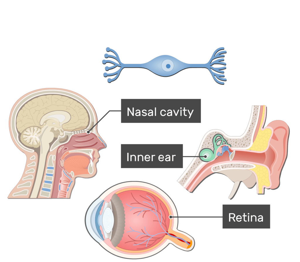
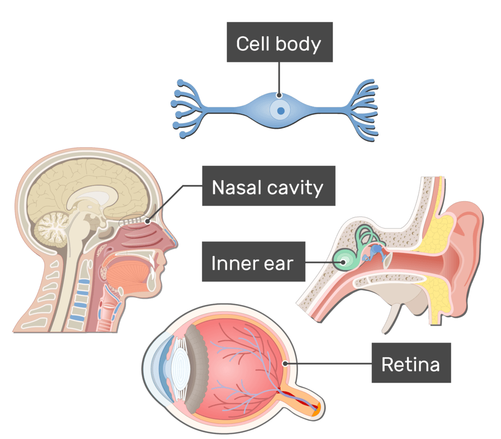
- One process serves as the axon and the other the dendrite. Both processes can have many smaller branches.
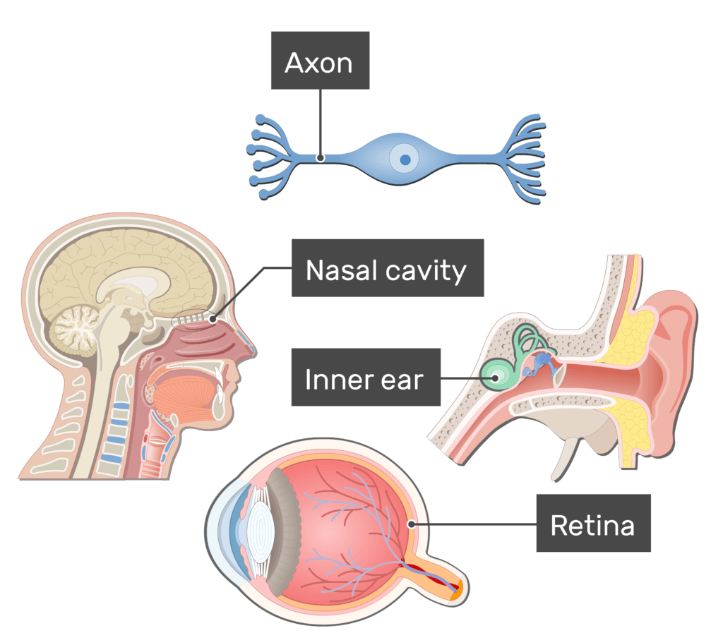

- Bipolar neurons are found in the retina of the eye, roof of the nasal cavity, and inner ear. They are always sensory and carry information about vision, olfaction, equilibrium, and hearing.
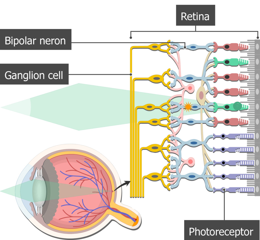
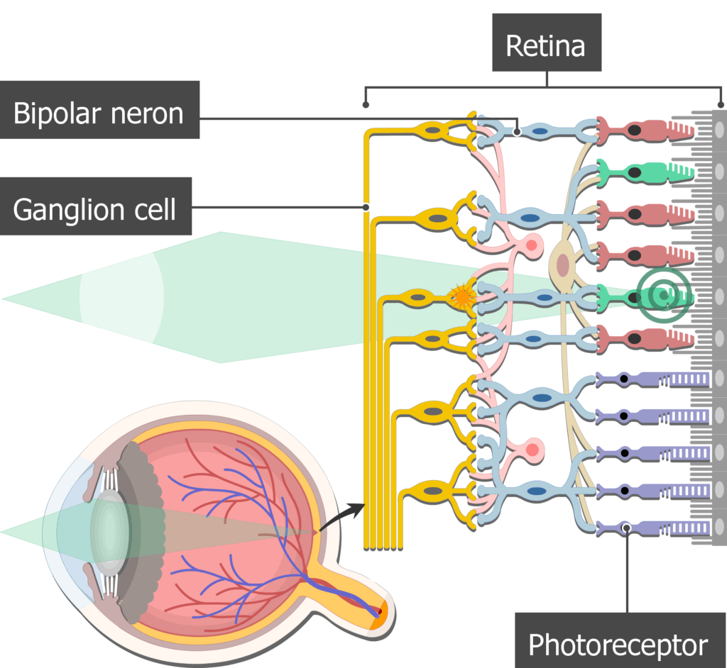
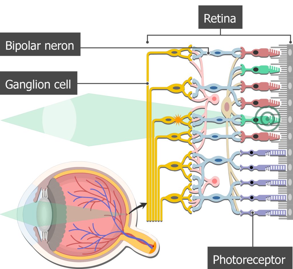
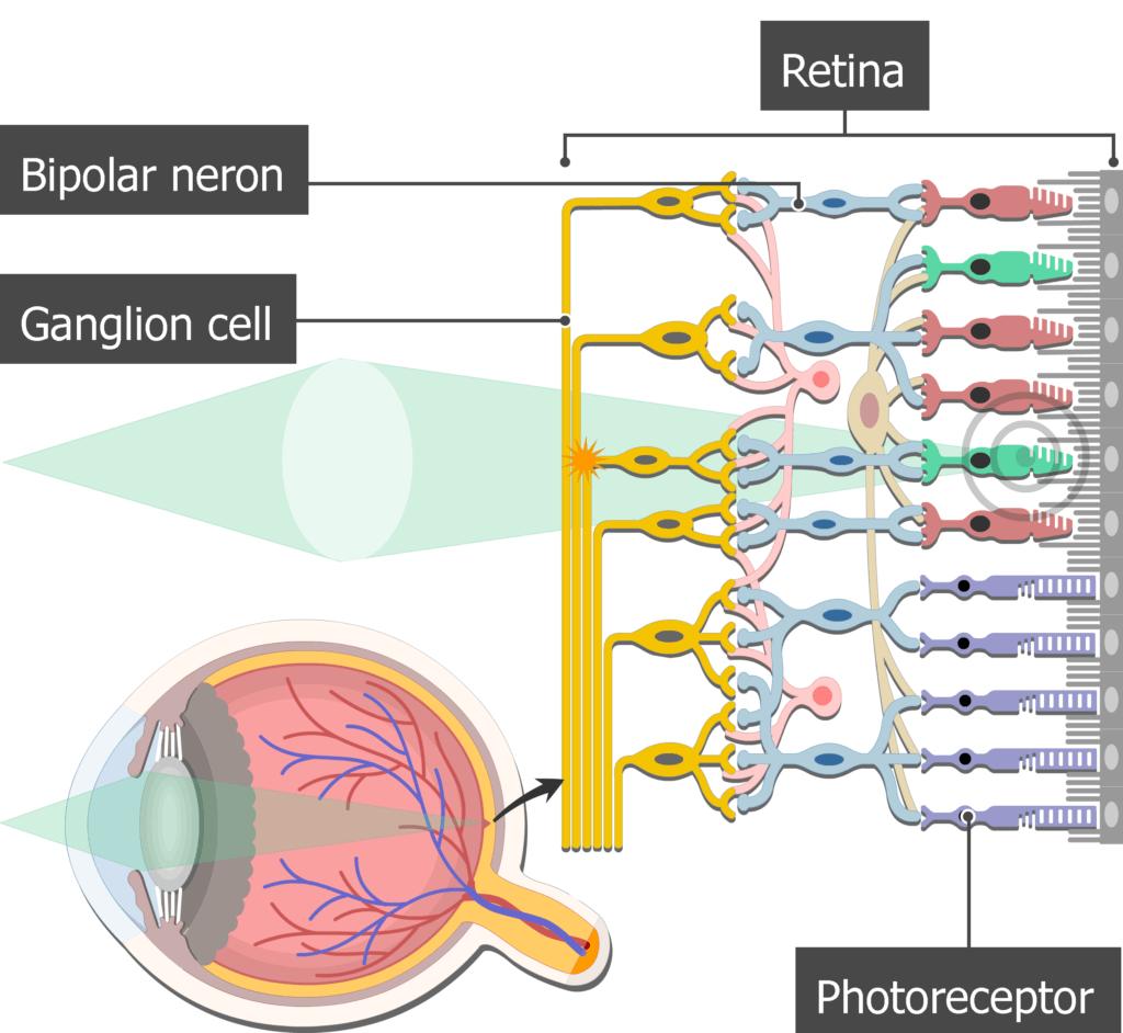
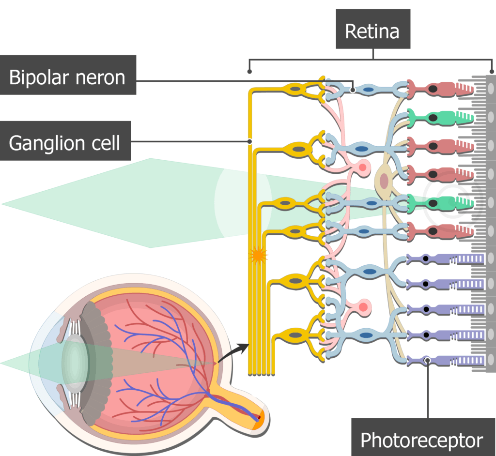
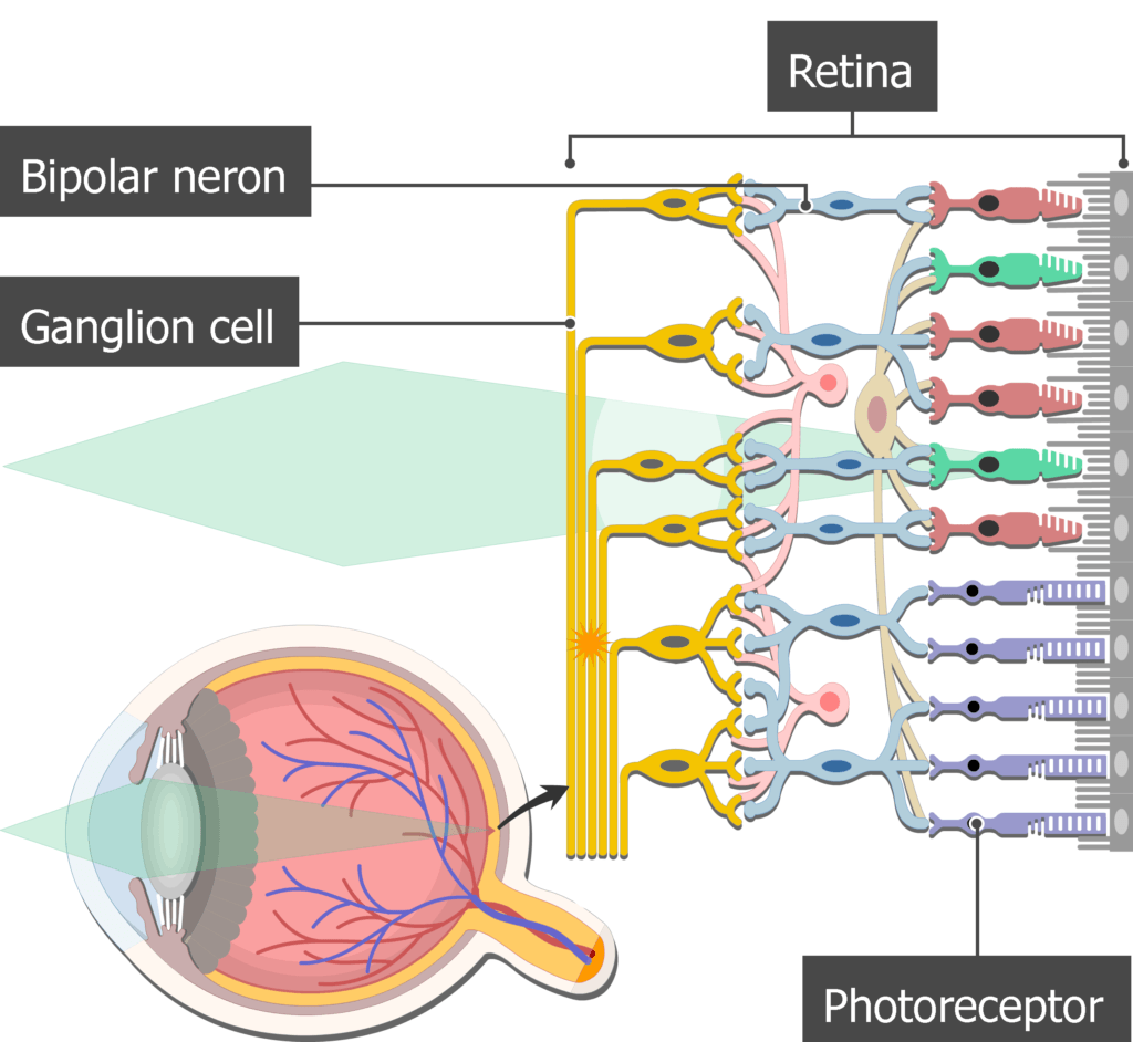
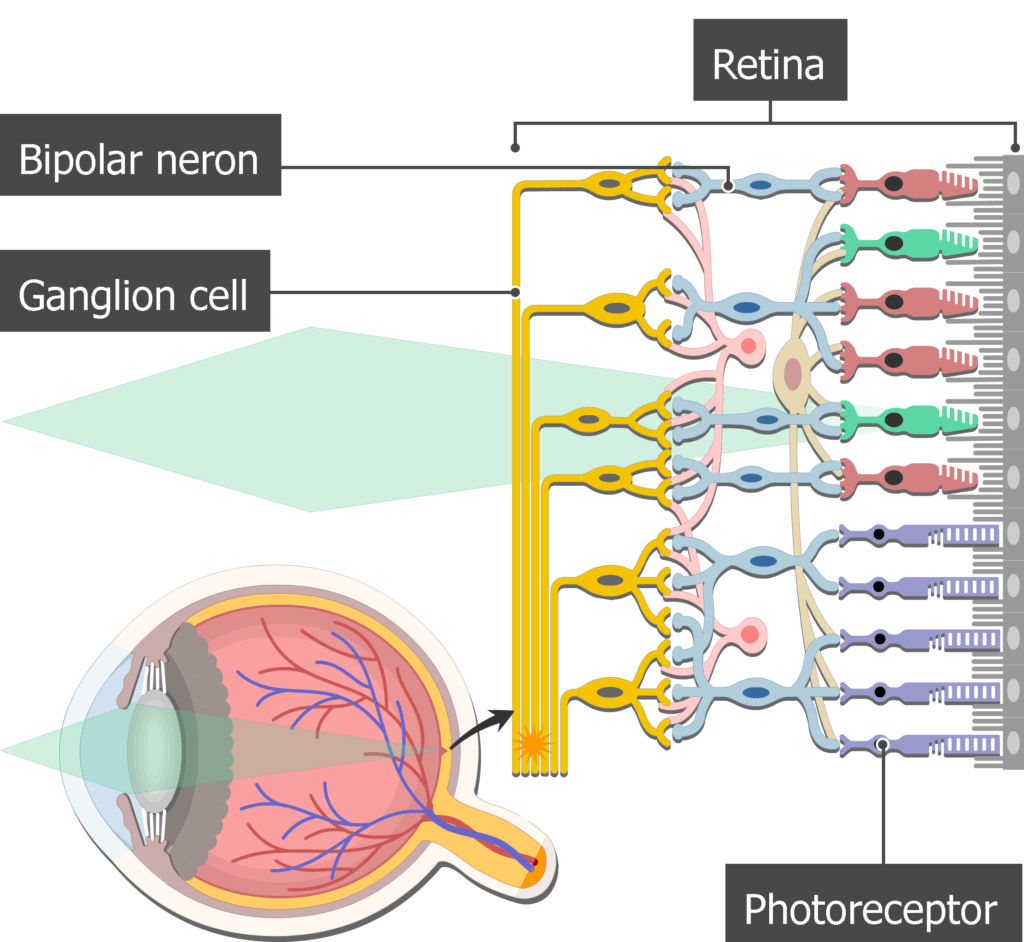
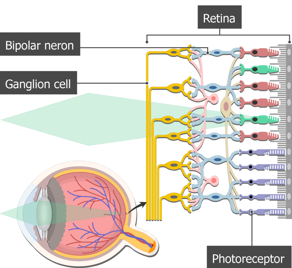
- In the eye, bipolar neurons form the middle layer of the retina.
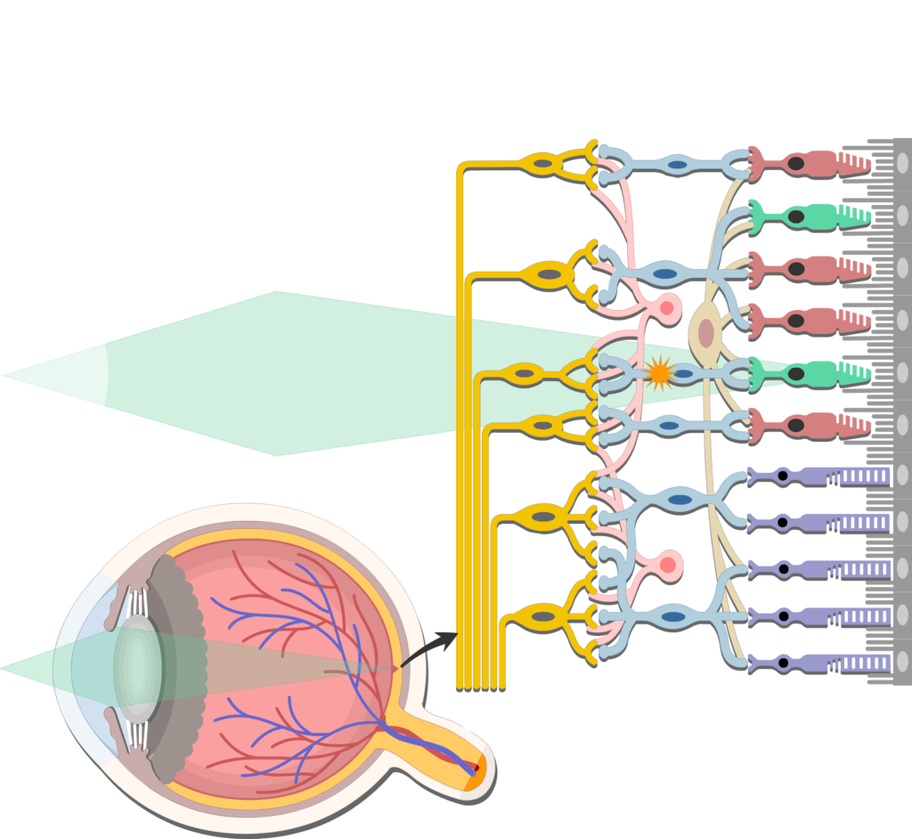

- Here they
 conduct impulses from photoreceptors (rods and cones) to ganglion cells.
- The ganglion cells then carry the visual signals to the brain via the optic nerve.
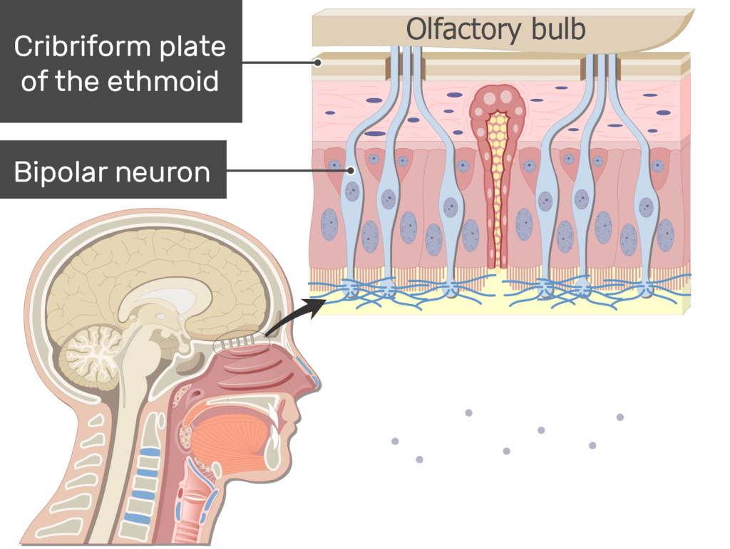
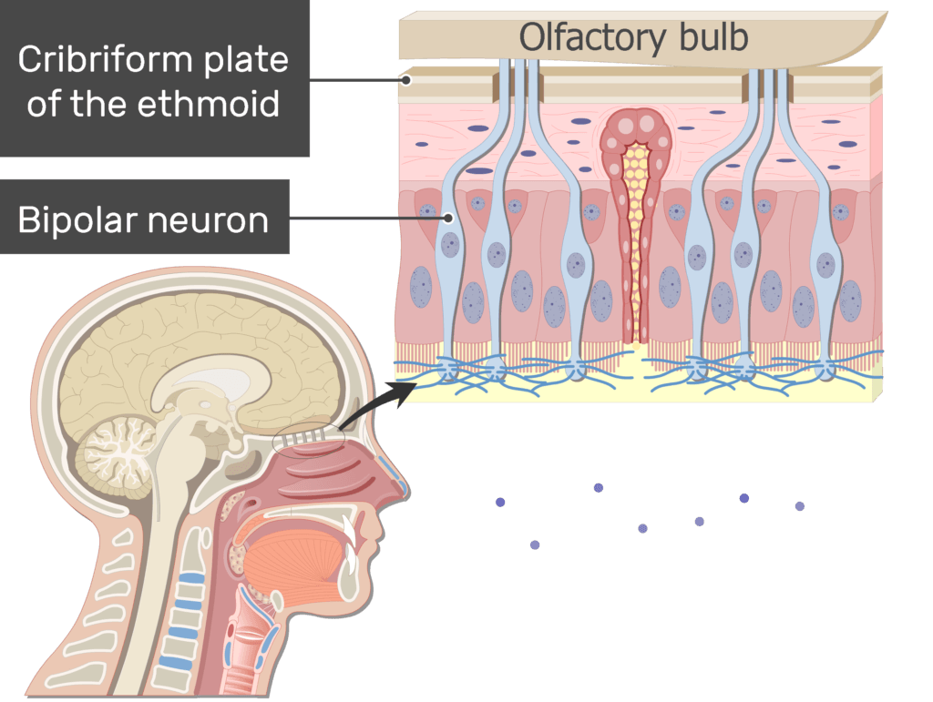
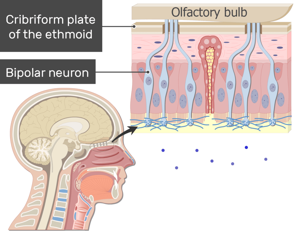
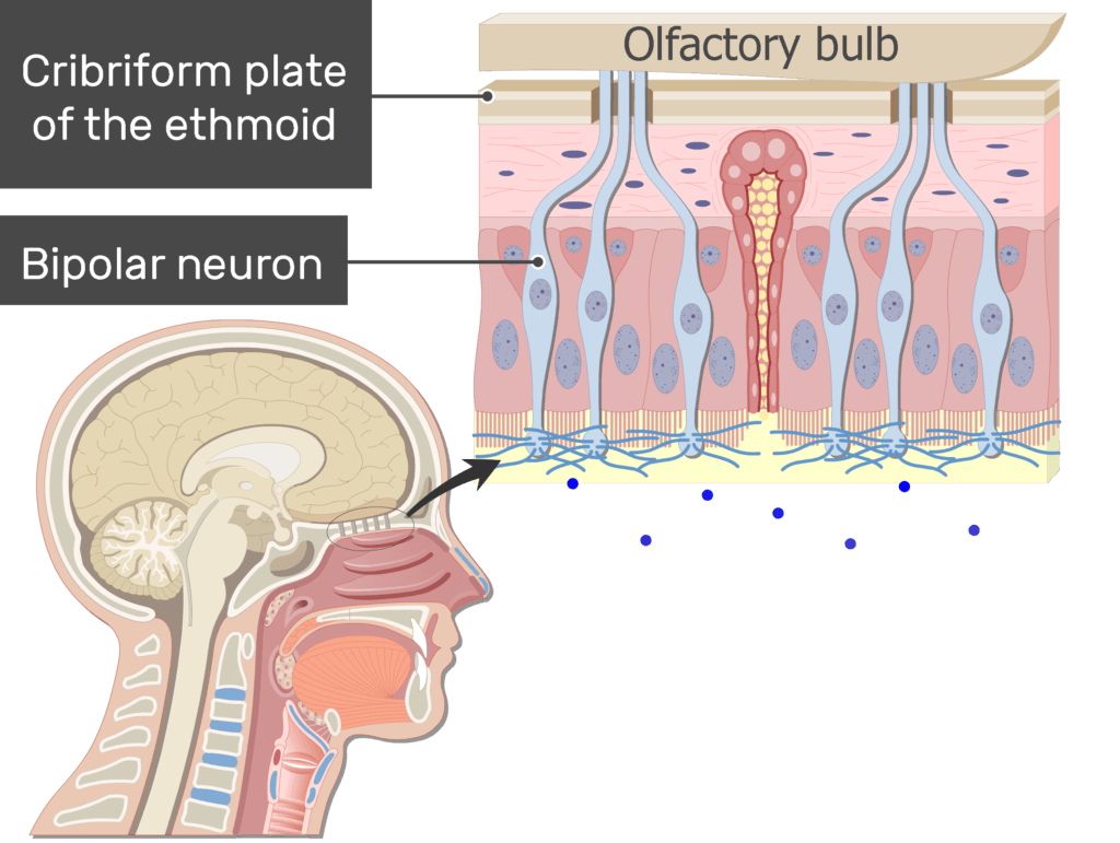
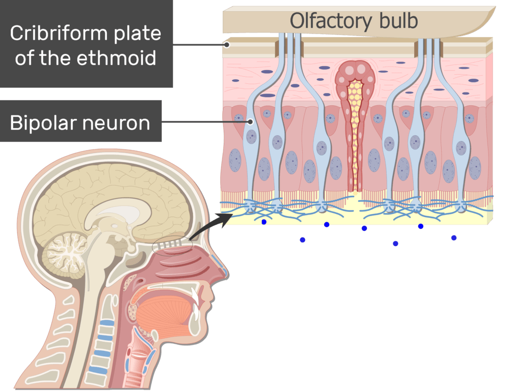
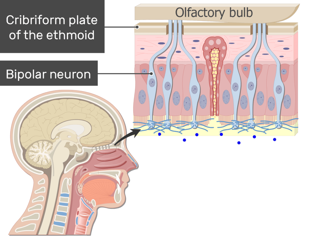
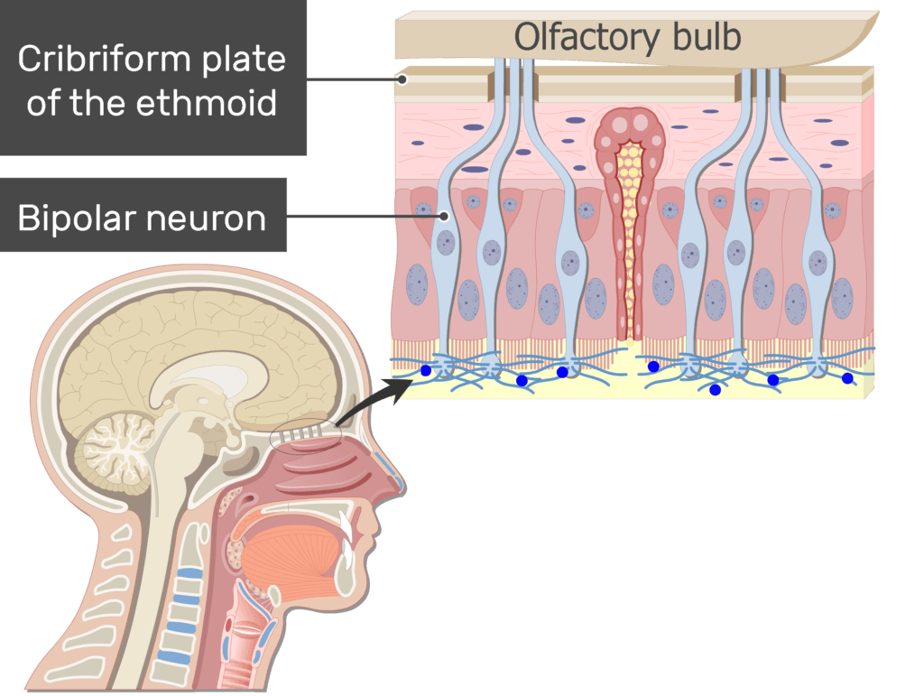
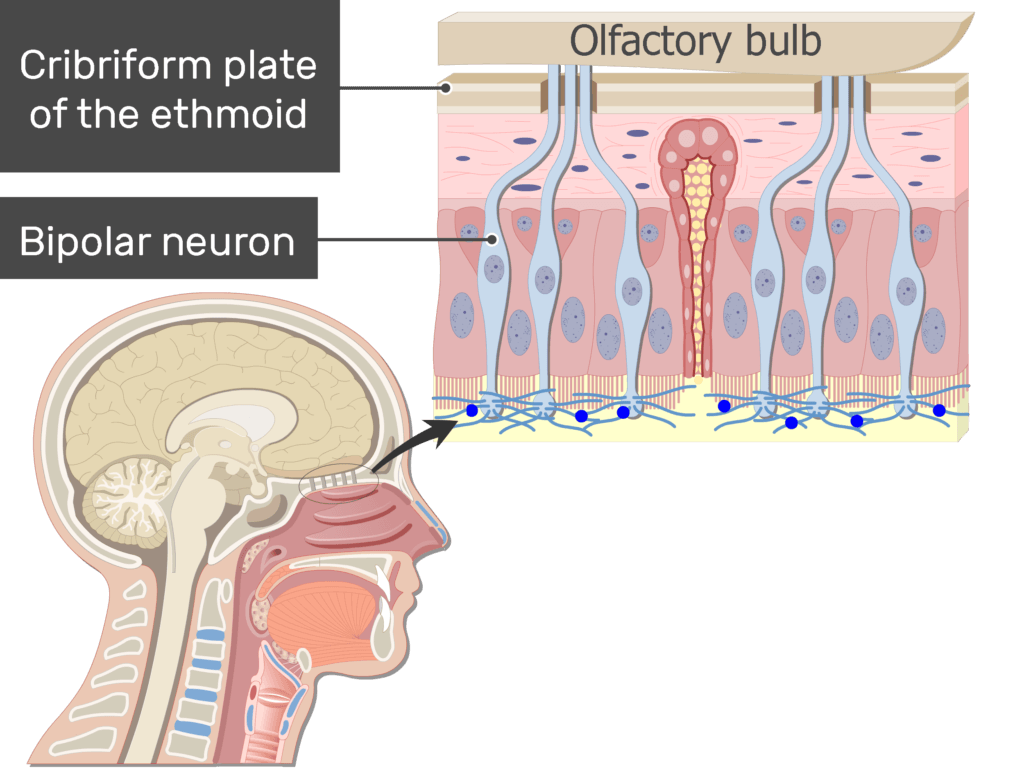
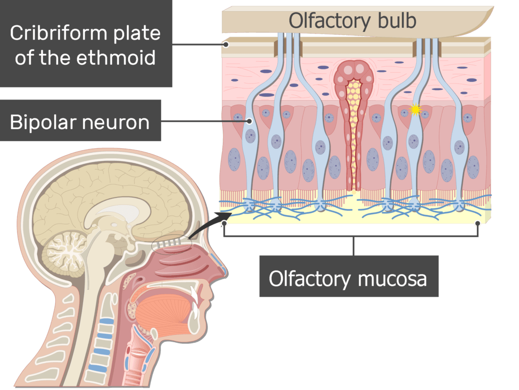
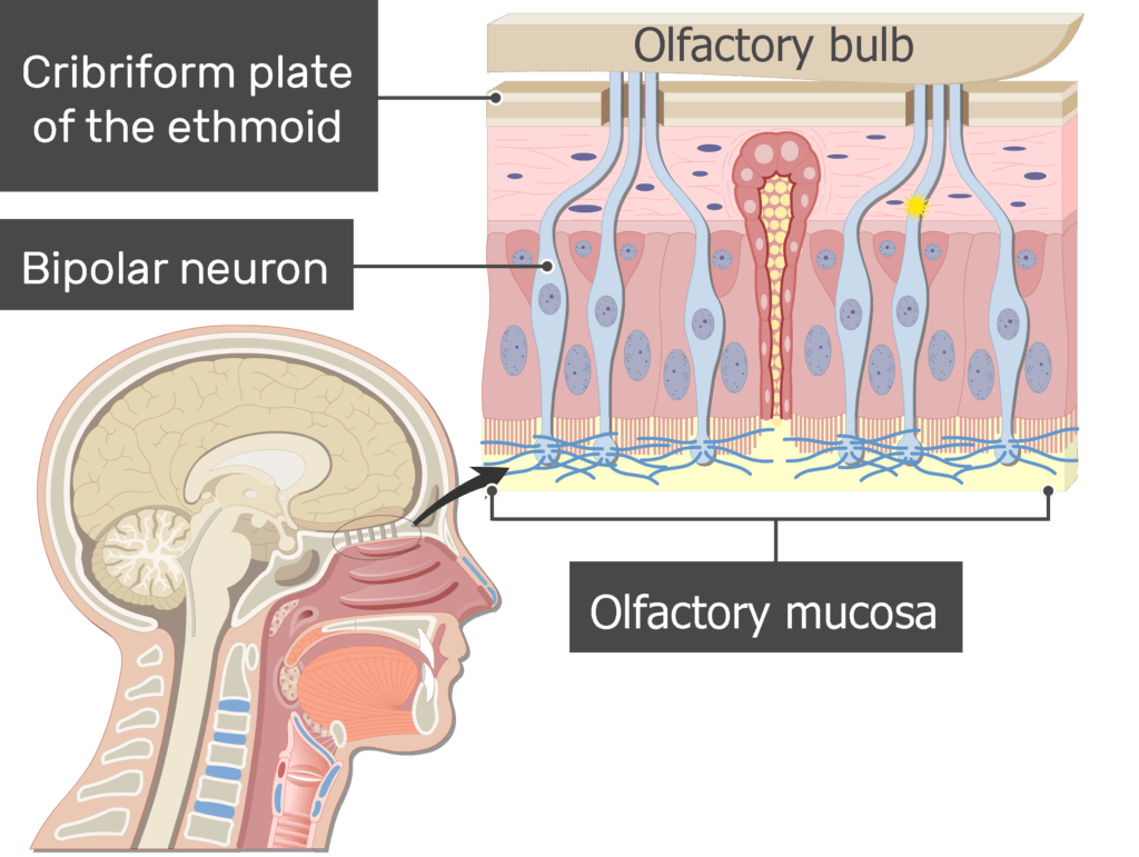
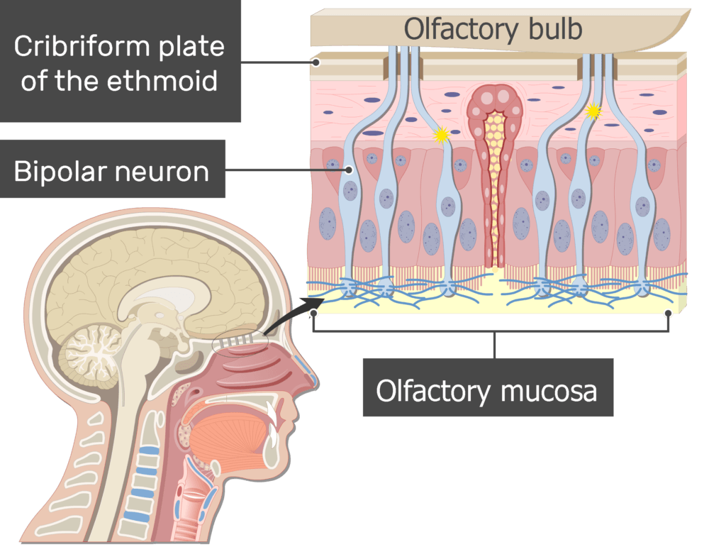
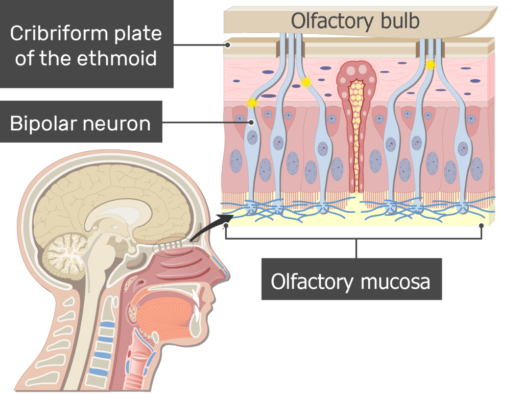
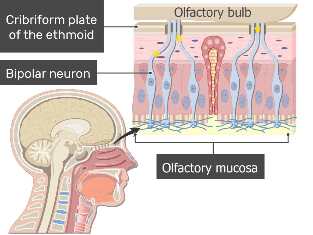
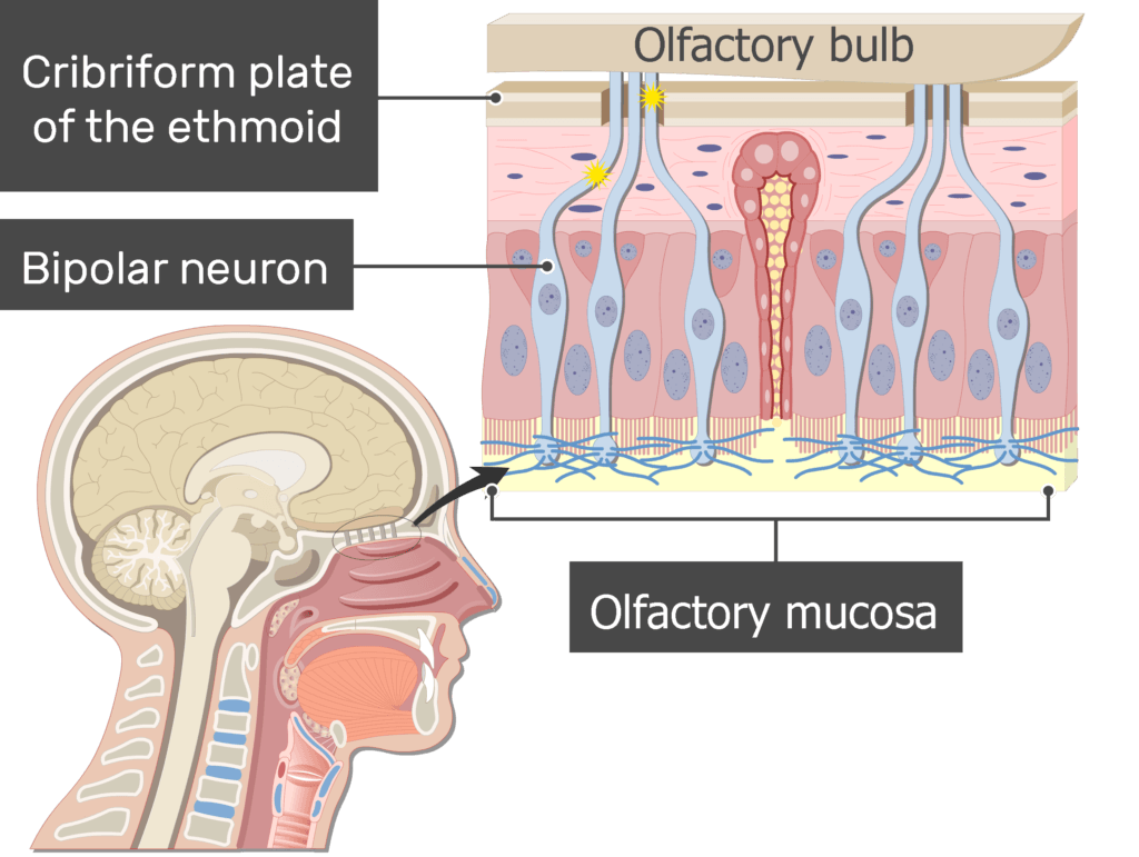
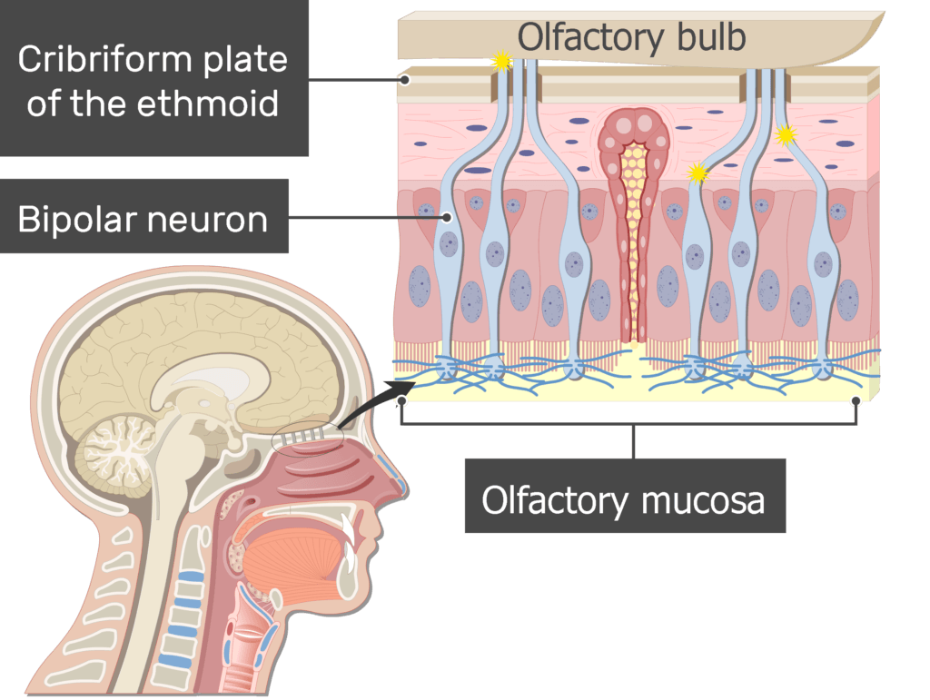
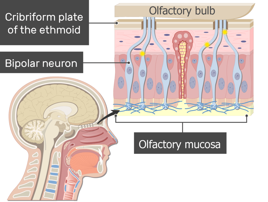
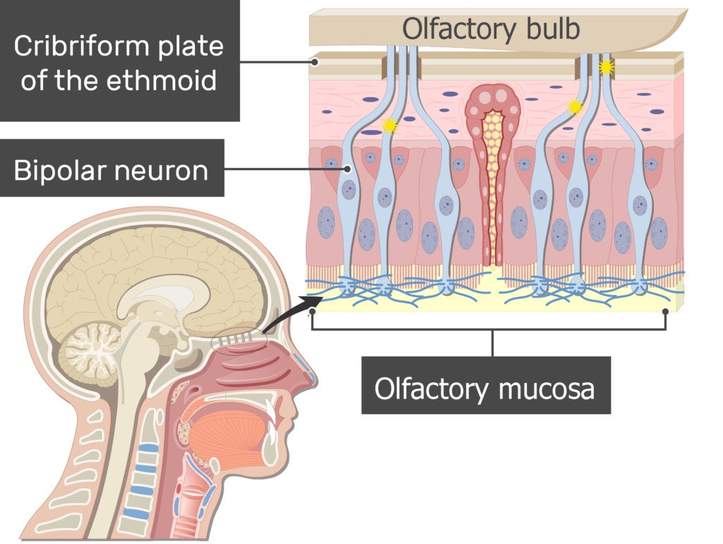
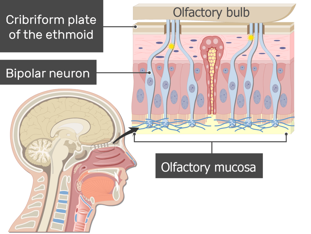
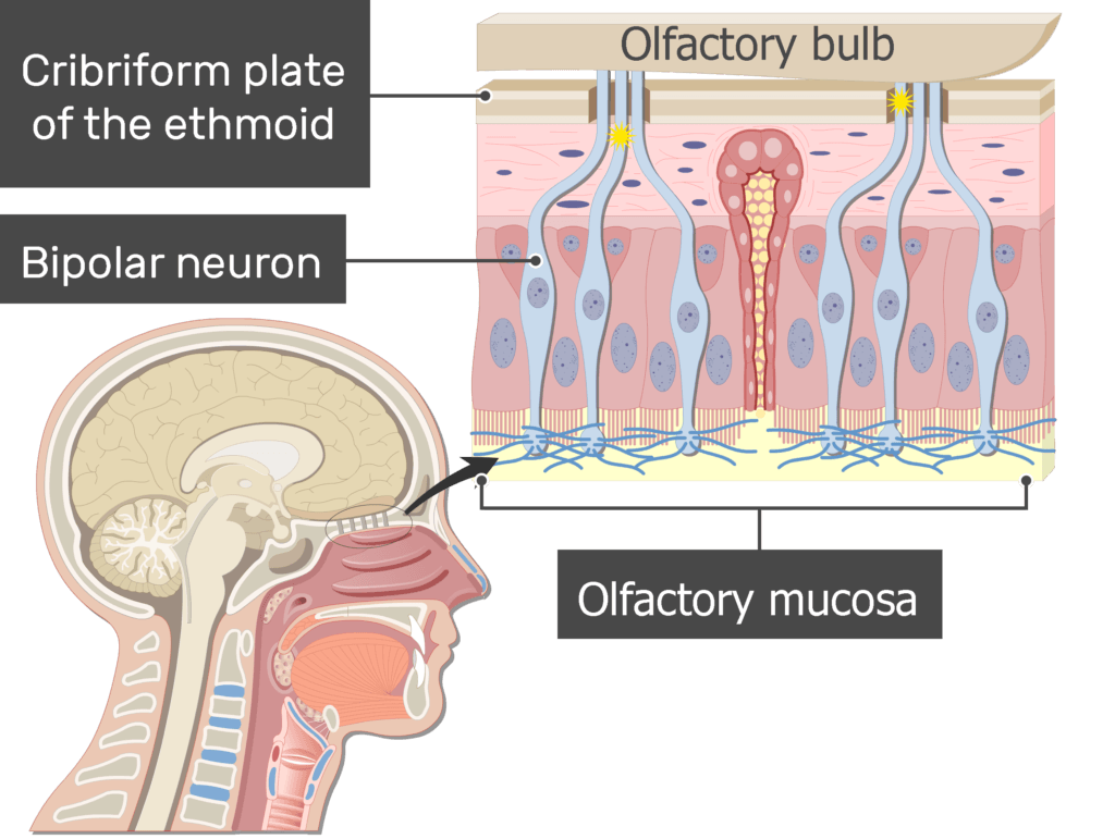
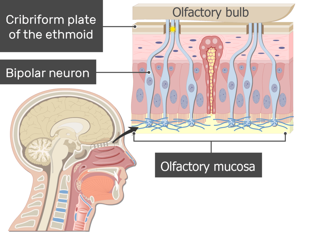
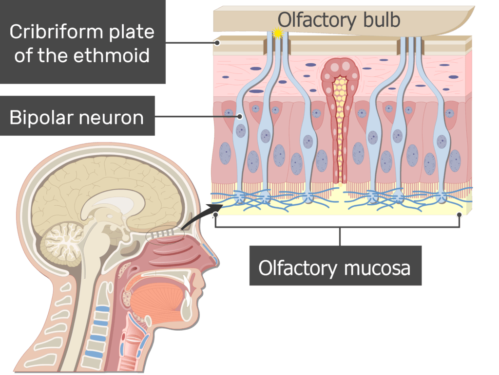
- The bipolar neurons in the nasal cavity are found in the olfactory mucosa where they serve as receptors.
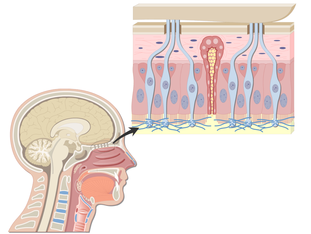
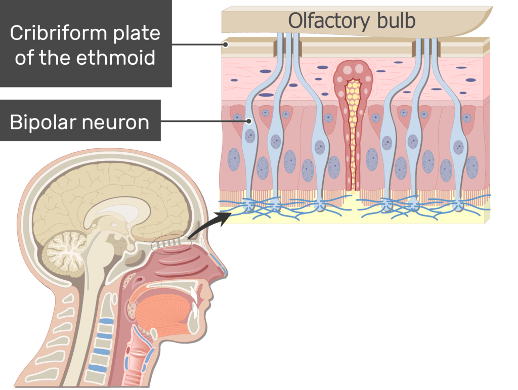
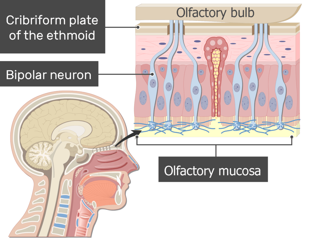
- When odorants are drawn into the nasal cavity, they bind to cilia that extend from the dendritic ends of the receptors.
Take your nervous system anatomy knowledge to the next level with these interactive quizzes and labeling exercises.
- In response, impulses are generated in the axons of the bipolar neurons. The axons carry the impulses through holes in the cribriform plate of the skull to the olfactory bulbs of the first cranial nerve.
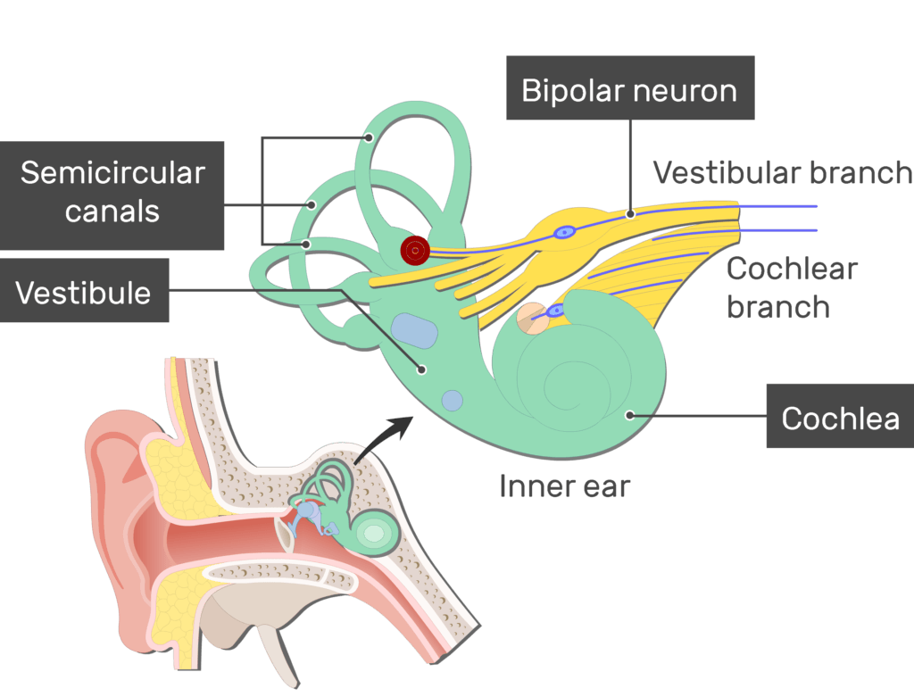
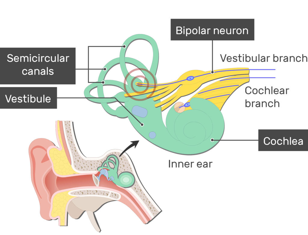
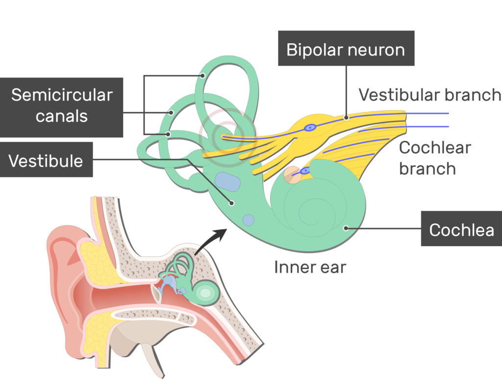
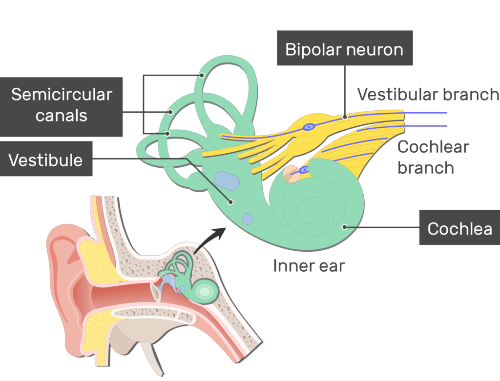
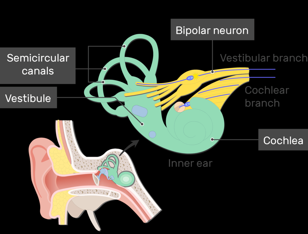
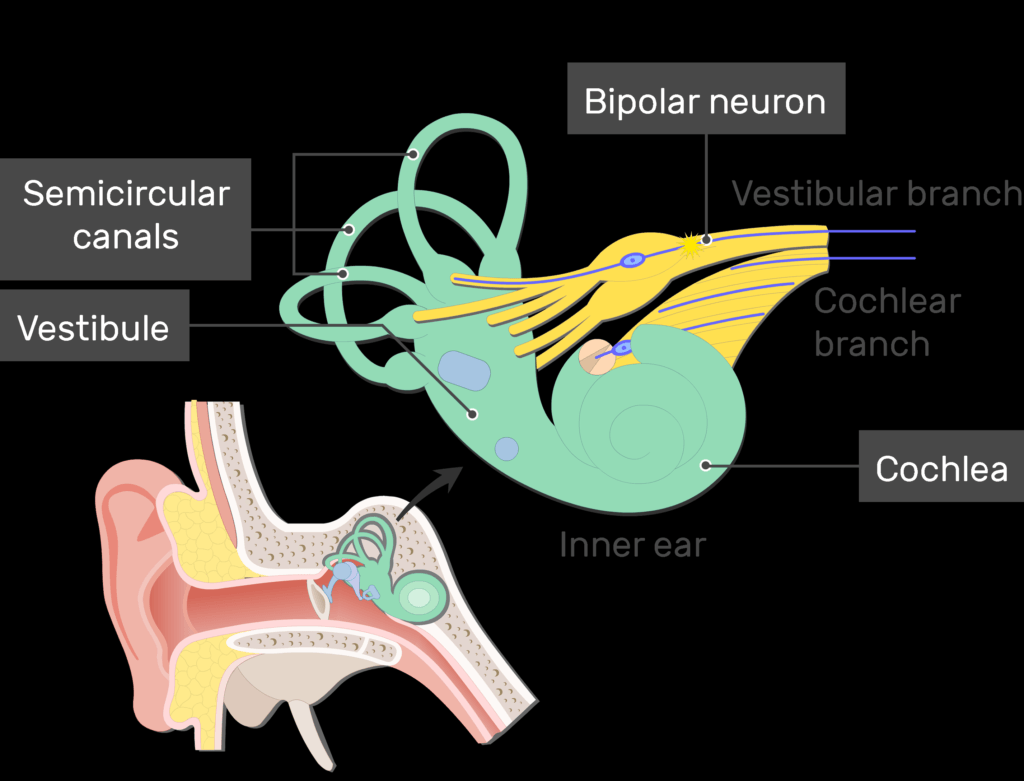
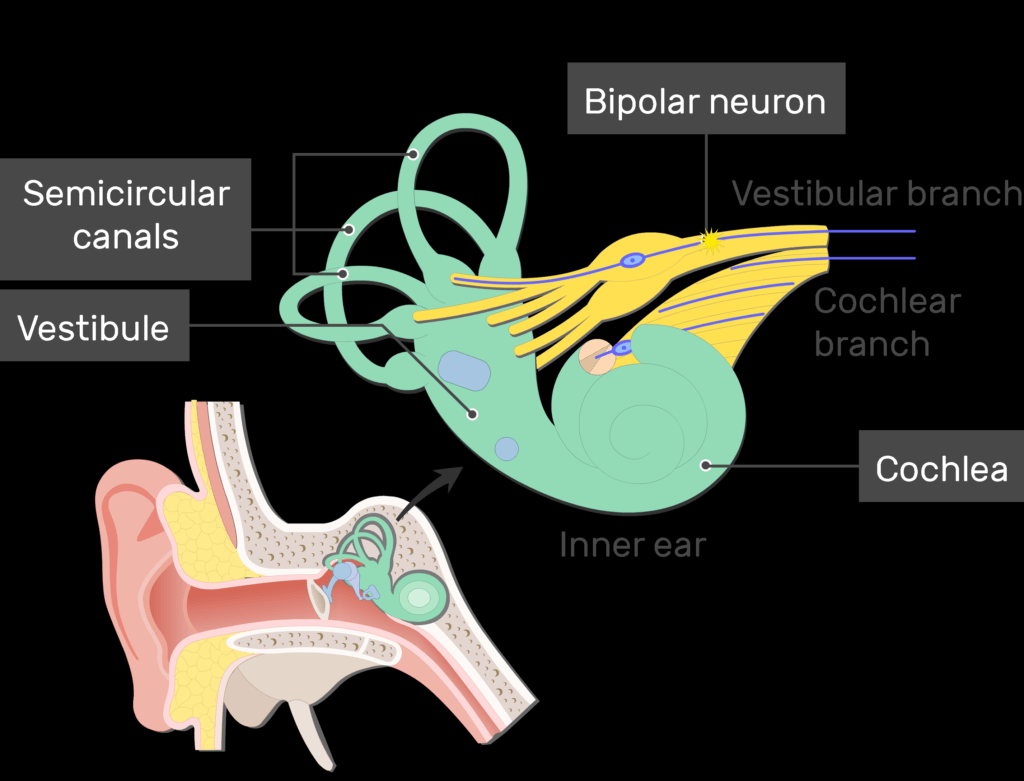
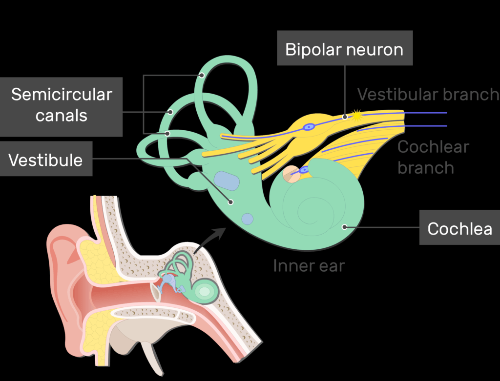
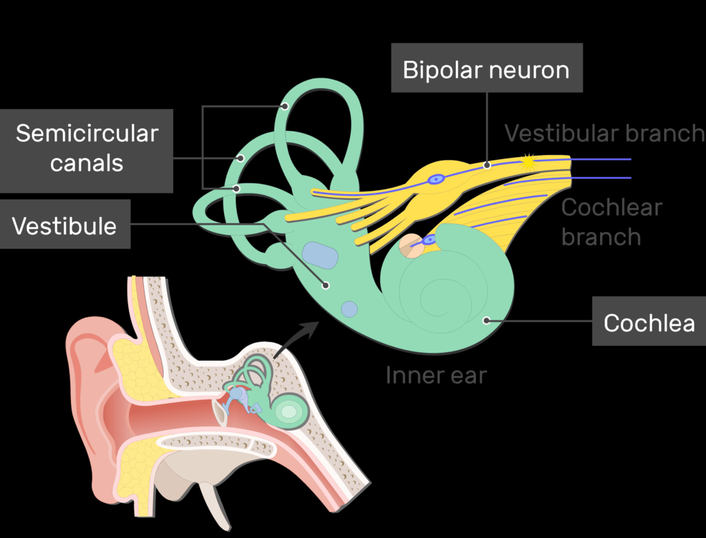
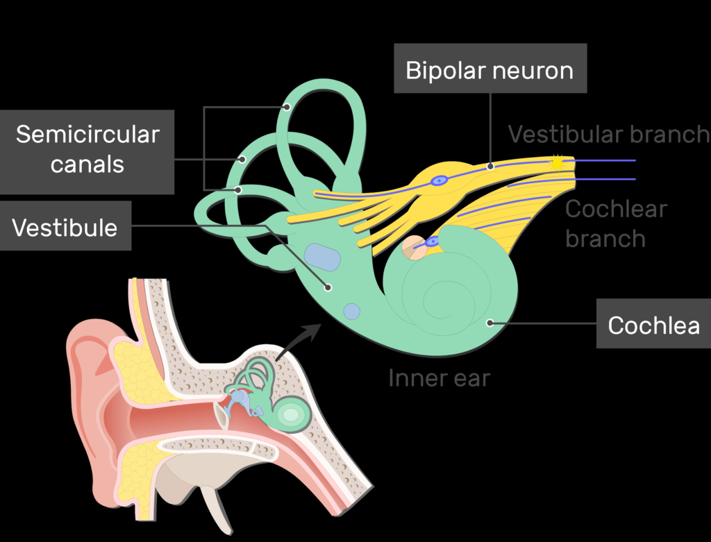
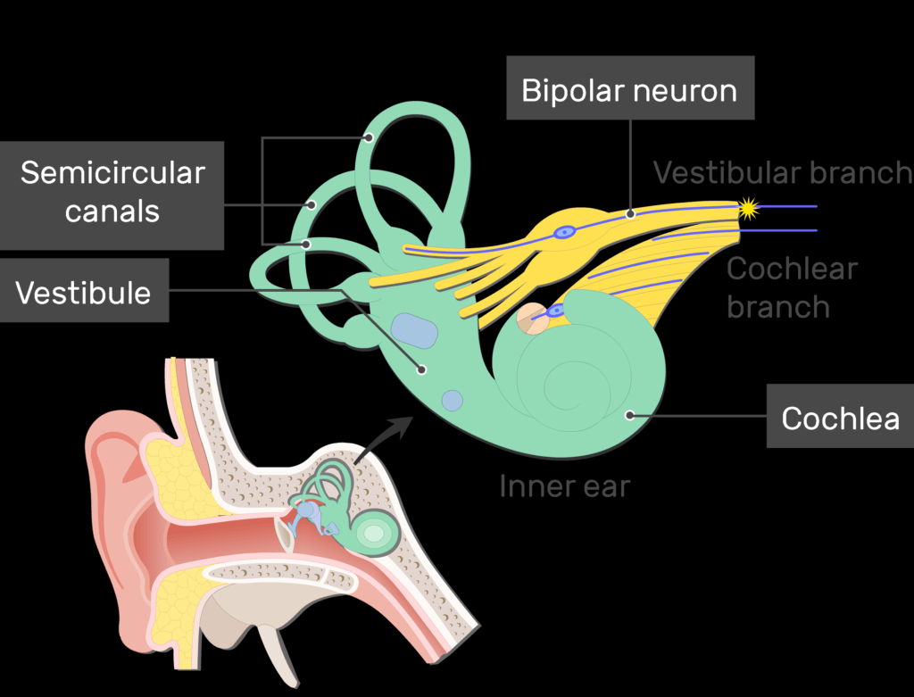
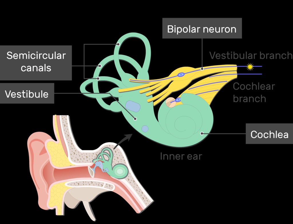
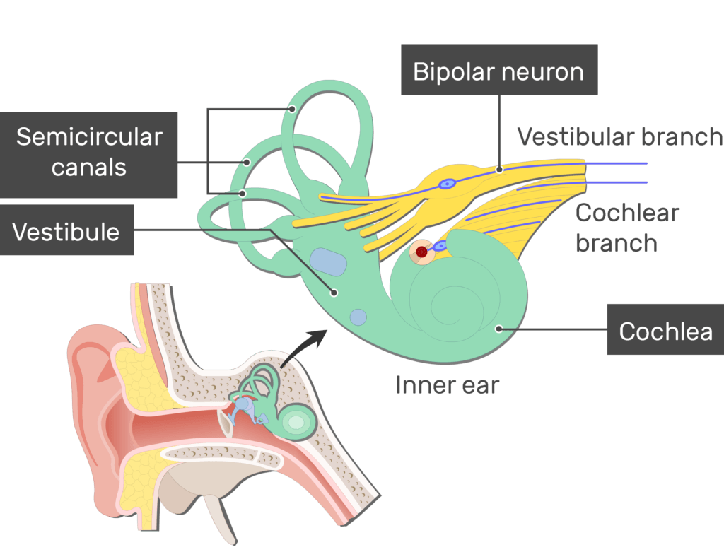
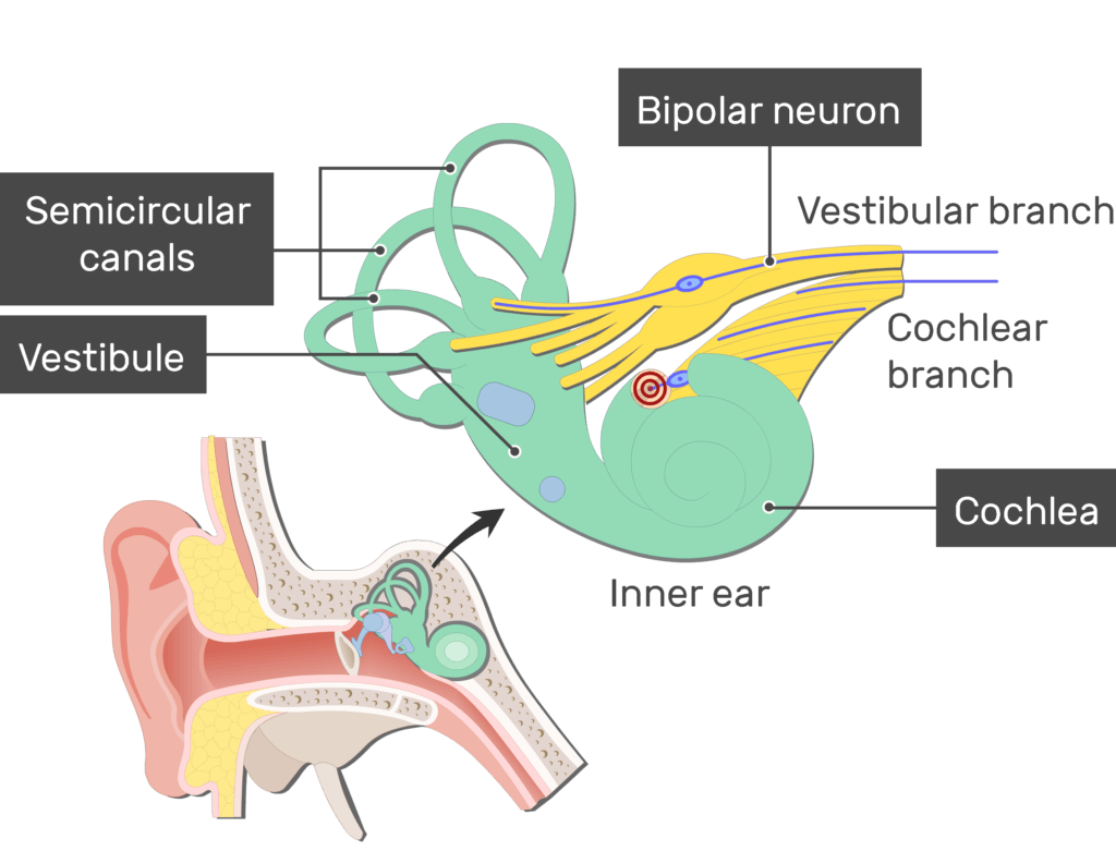
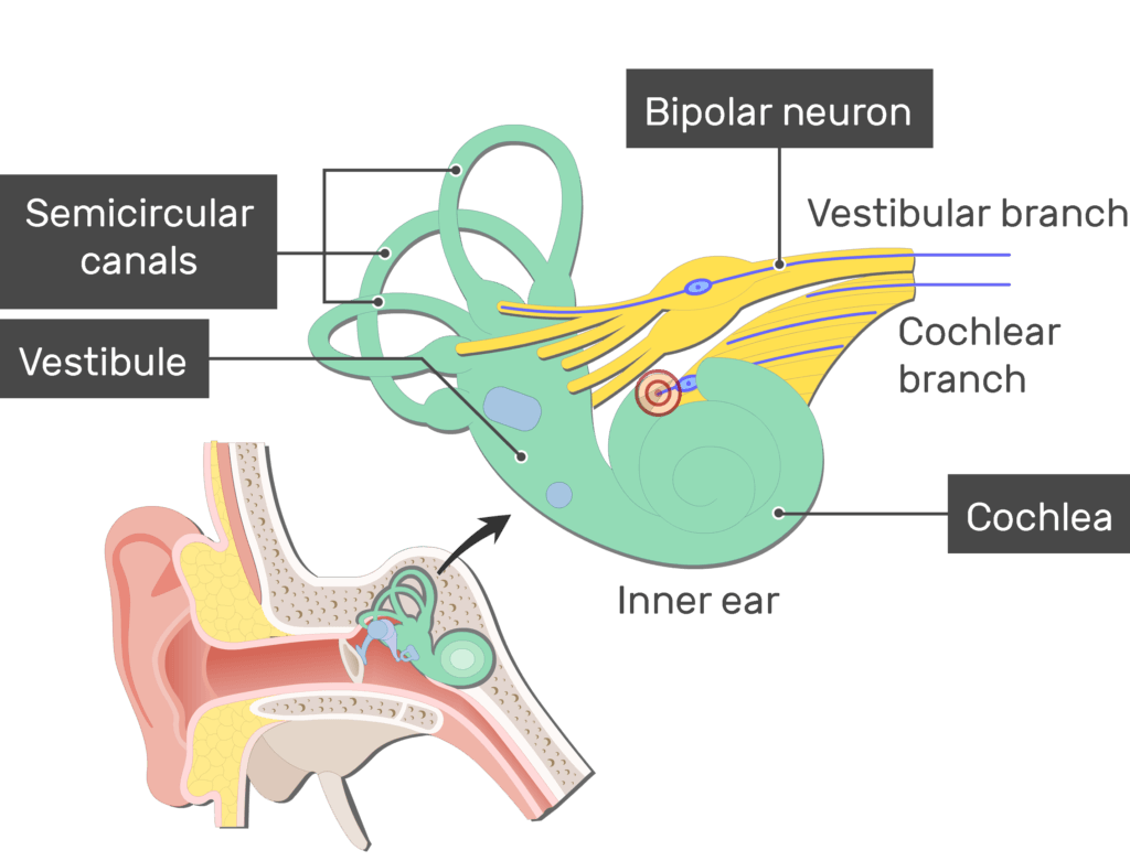
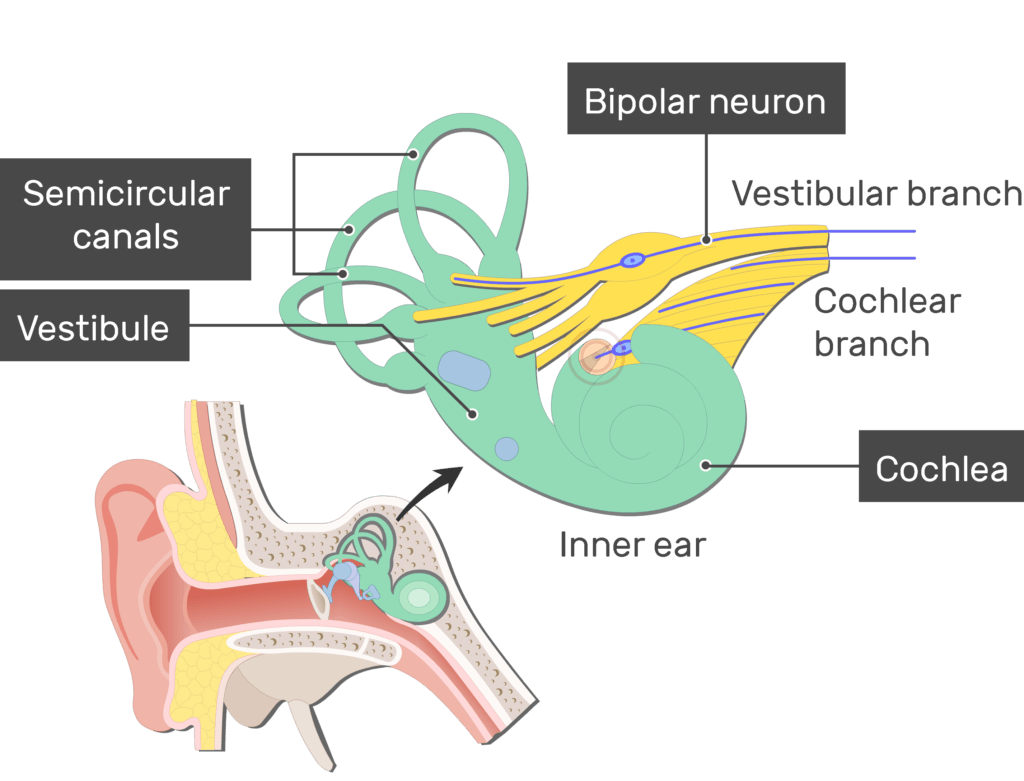
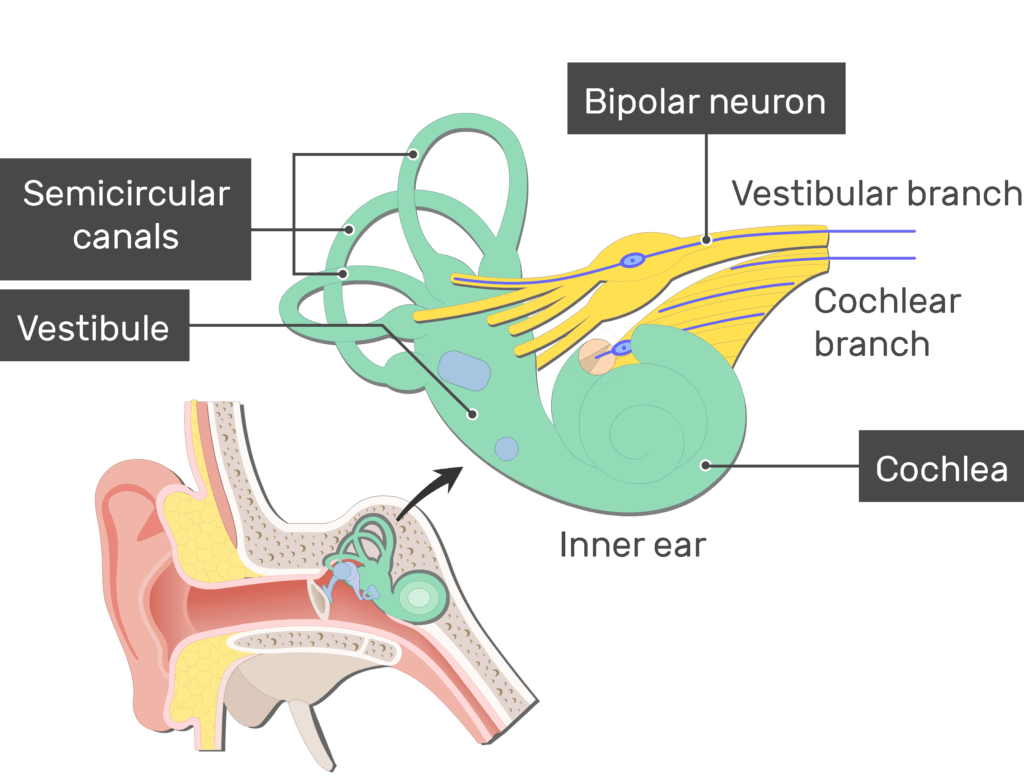
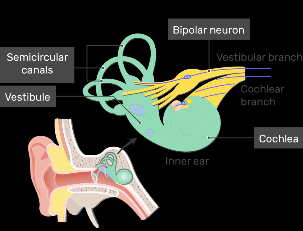
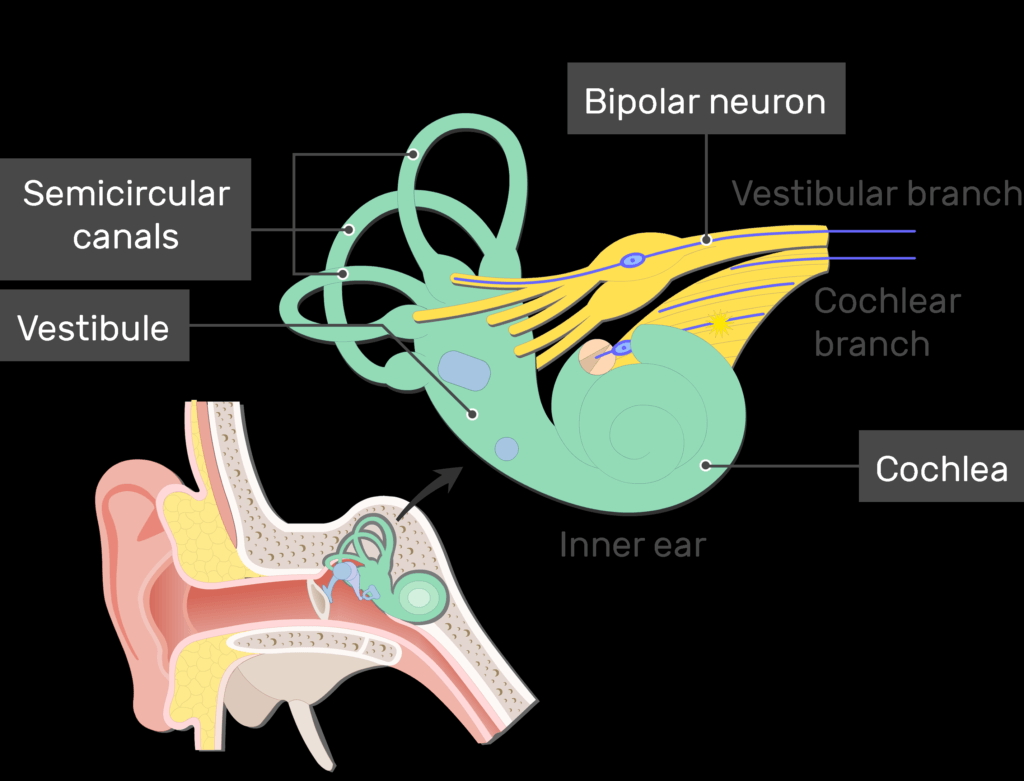
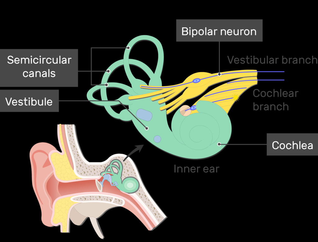
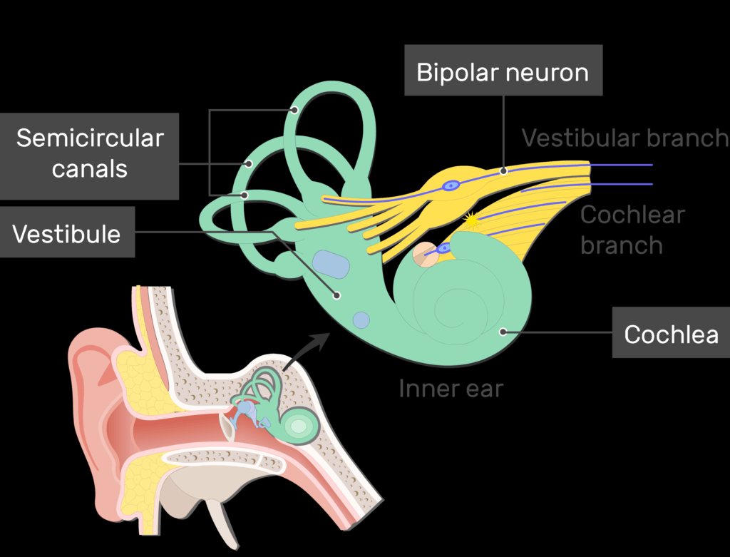
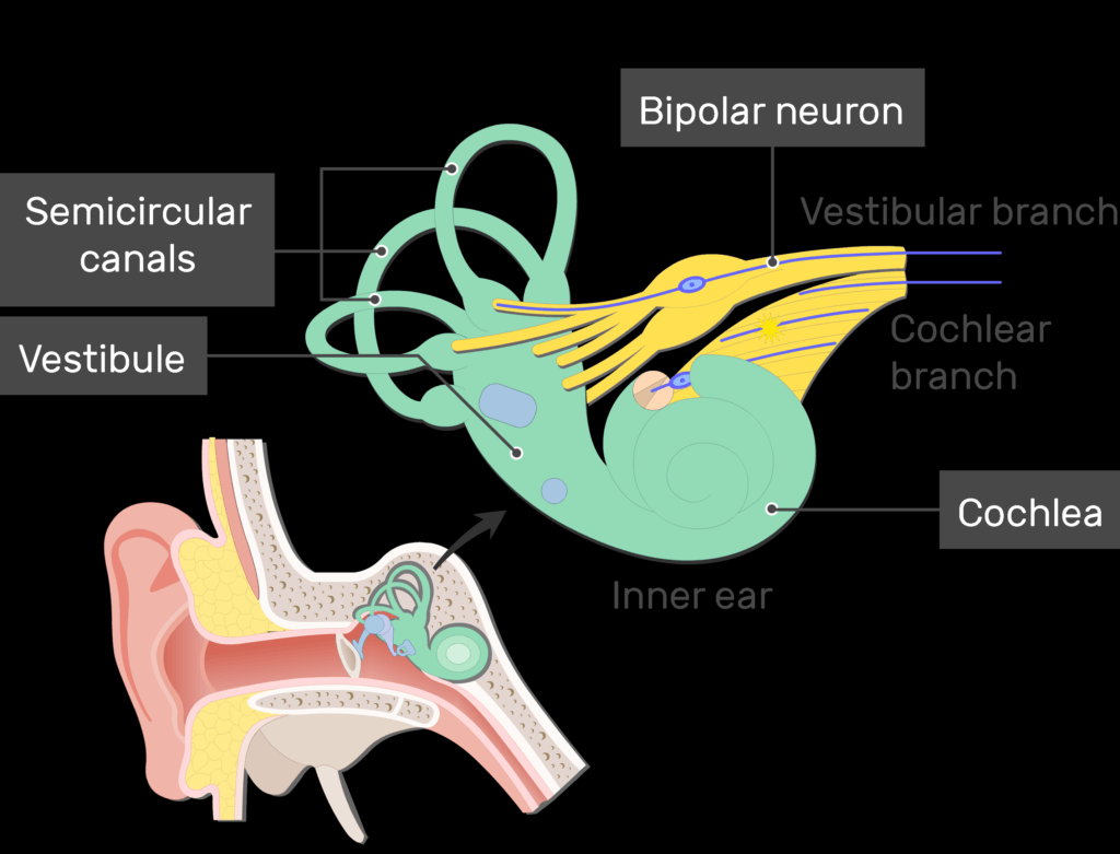
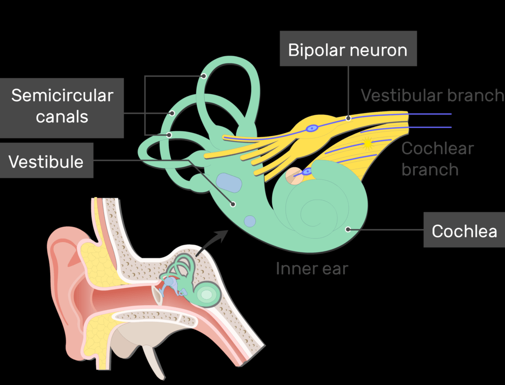
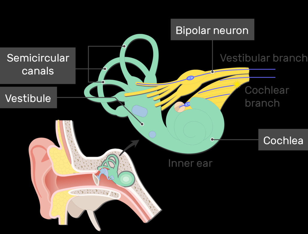
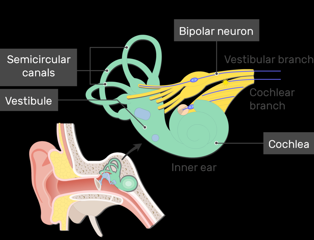
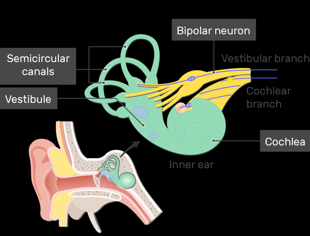
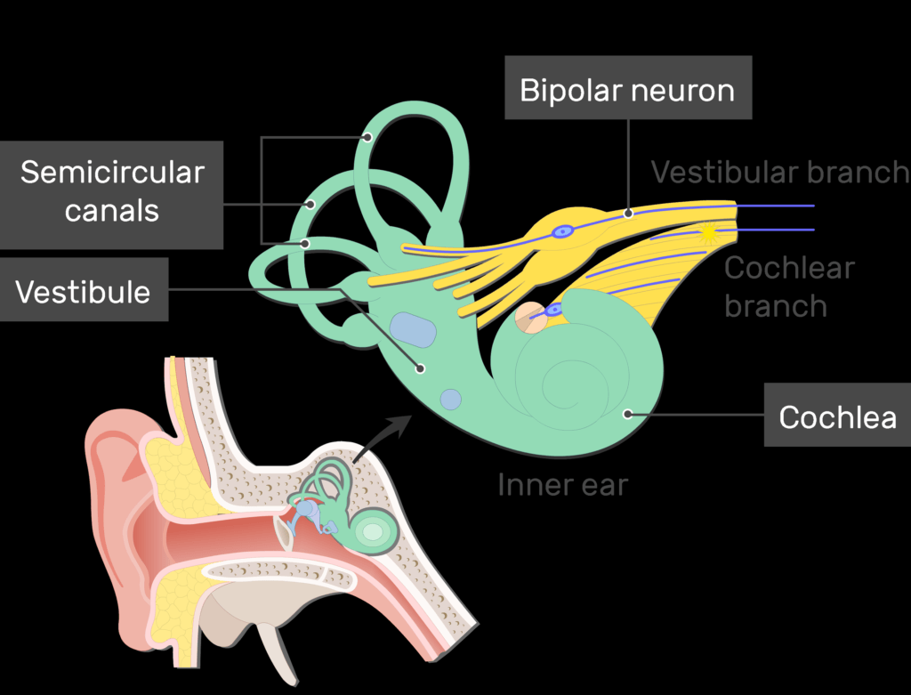
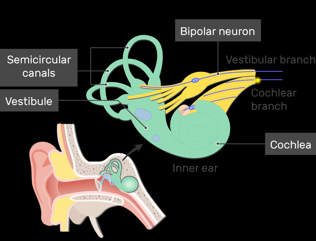
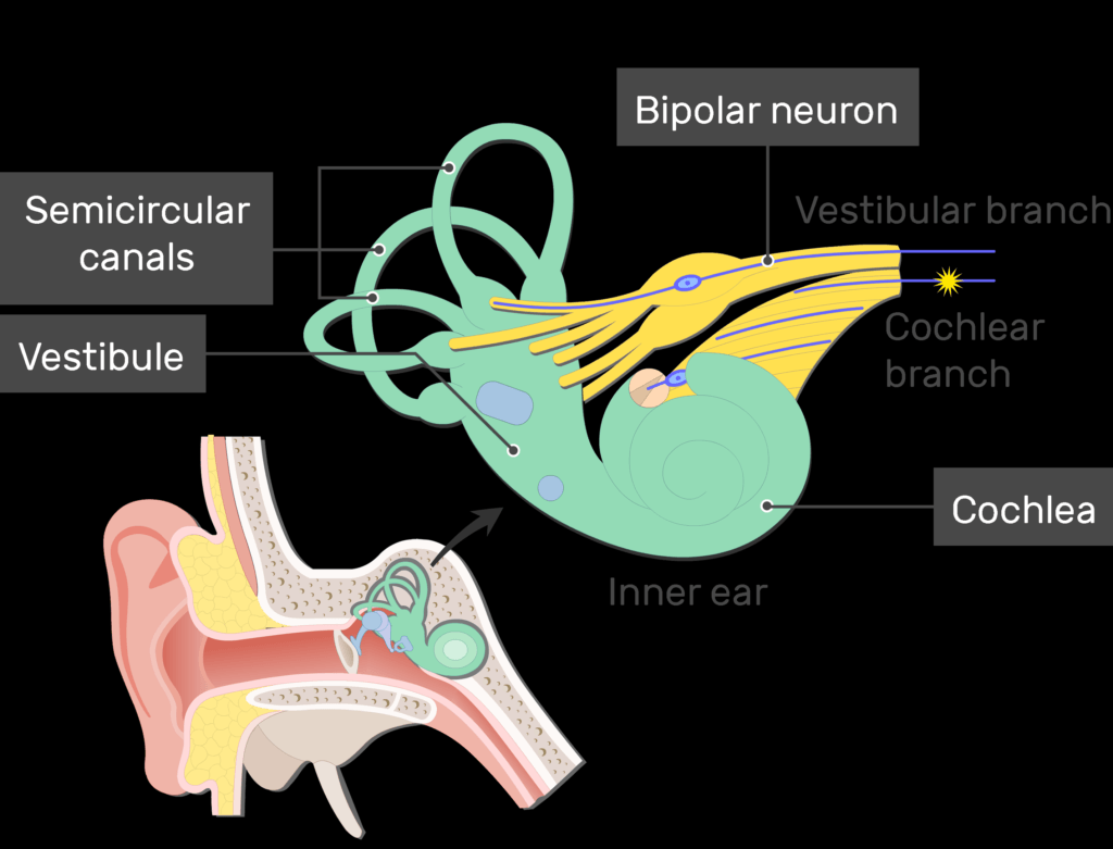
- In the inner ear, bipolar neurons make up the vestibular and cochlear branches of the vestibulocochlear nerve (cranial nerve VIII).
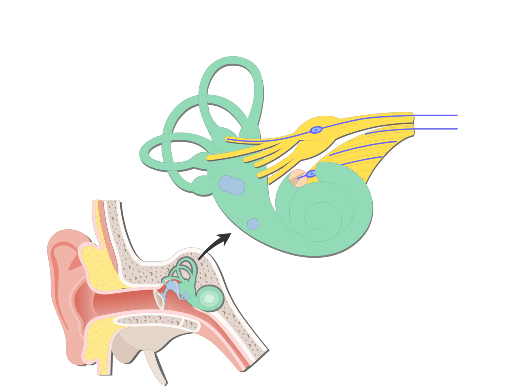

- The vestibular branch carries signals into brain about balance (equilibrium).
- The cochlear branch carries signals about hearing.
Related Articles
Unipolar Neuron – Structure and Functions
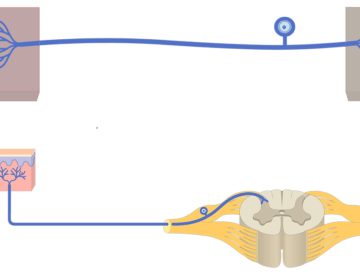
Unipolar (pseudounipolar) neurons have one process that attached to the cell body.

Types of Neurons
Classification of Neurons; explained beautifully in an illustrated and interactive way.
Neuron: structure, function and diagram
Neuron (Basic Structure and Functions); explained beautifully in an illustrated and interactive way.