



Splenius Capitis Muscle
Skeletal muscle contraction physiology
Last update:
Excitation-contraction coupling
Every skeletal muscle contraction begins with the release of neurotransmitter acetylcholine from the nerve that innervates the muscle. Acetylcholine causes electrical changes on the plasma membrane of the muscle cells, which in turn trigger a series of events within the muscle cell itself, which eventually lead to the muscle fiber contraction.
This fast sequence of events between the electrical changes of the membrane and muscle contraction is described with a physiological term excitation-contraction coupling, or shortened ec coupling. To further simplify the meaning of this term, it refers to the coupling (connecting) of the process of excitation of the cell membrane and consequential muscle contraction.
Mechanism of muscle contraction and ec coupling
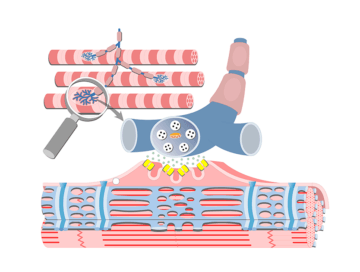
The excitation process begins when the acetylcholine (ACh) is released by a motor neuron at the neuromuscular junction.
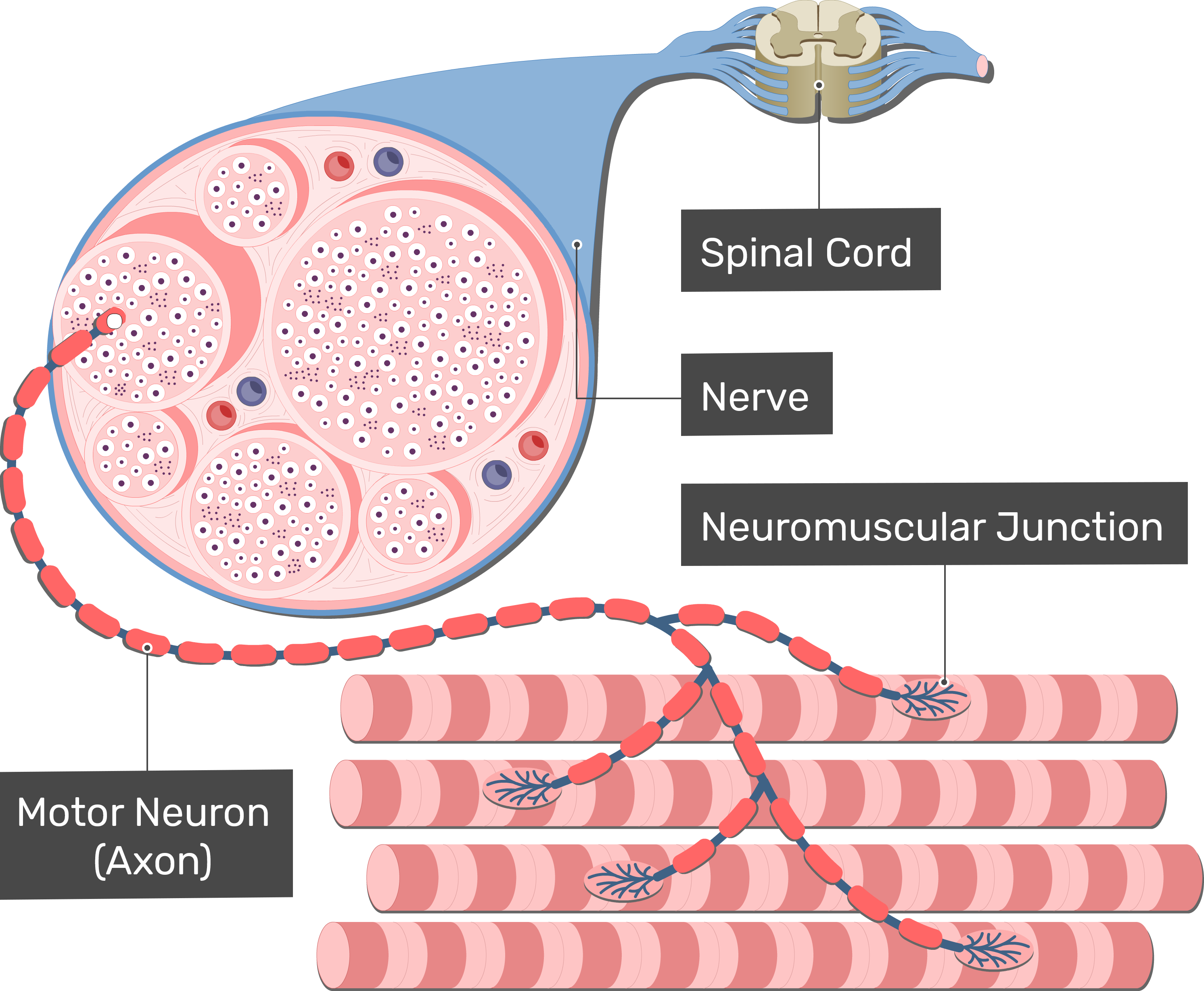
Once the Ach binds to the muscle cell membrane (sarcolemma), it causes the ACh-gated sodium channels to open. Then, large amounts of sodium (Na+) enter the muscle cell. Large influx of sodium leads to depolarization of that specific part of the cell membrane. Local change of electrical gradient causes the opening of voltage-gated sodium channels, which further increases the sodium influx and triggers an action potential across the whole muscle cell membrane.
This action potential propagates along the cell membrane and causes the sarcoplasmic reticulum to release large amounts of calcium ions (Ca++) into the cell. Remember, sarcoplasmic reticulum of a skeletal muscle cell is an equivalent to endoplasmic reticulum in other cells and its main feature in the muscle cell is to store ions of calcium.
The ions of calcium which are now in the cell initiate the attraction between the  actin and myosin filaments. The filaments then slide along each other, which causes the shortening of the muscle fibers, or in other words – muscle contraction.
When the muscle cell stimulation ends, the ions of calcium are pushed back into the sarcoplasmic reticulum. They remain stored within it until the next muscle contraction.


Actin and myosin filaments shown sliding one alongside the other.
Test yourself
Contraction Physiology
References
- Betts, J. G., Young, K. A., Wise, J. A., Johnson, E., Poe, B., & Kruse, D. H. (2022). Anatomy and Physiology (2nd ed.). OpenStax. https://openstax.org/details/books/anatomy-and-physiology-2e
- Hall, J. E., & Guyton, A. C. (2016). Guyton and Hall Textbook of Medical Physiology (13th ed.). Elsevier, Philadelphia PA
- Calderón, J. C., Bolaños, P., & Caputo, C. (2014). The excitation–contraction coupling mechanism in skeletal muscle. Biophysical Reviews, 6(1), 133–160. https://doi.org/10.1007/s12551-013-0135-x
