


Major system arteries of the body
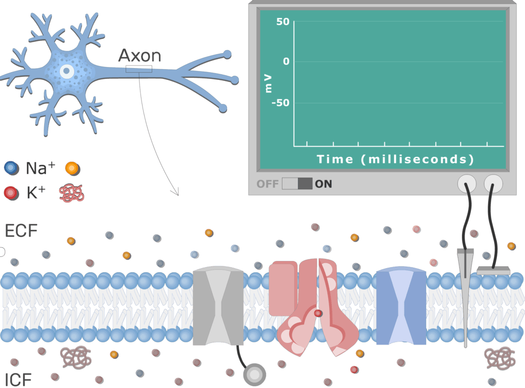
Neuron Action Potential Sequence of Events
Last update:
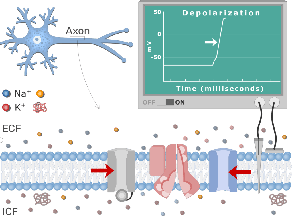
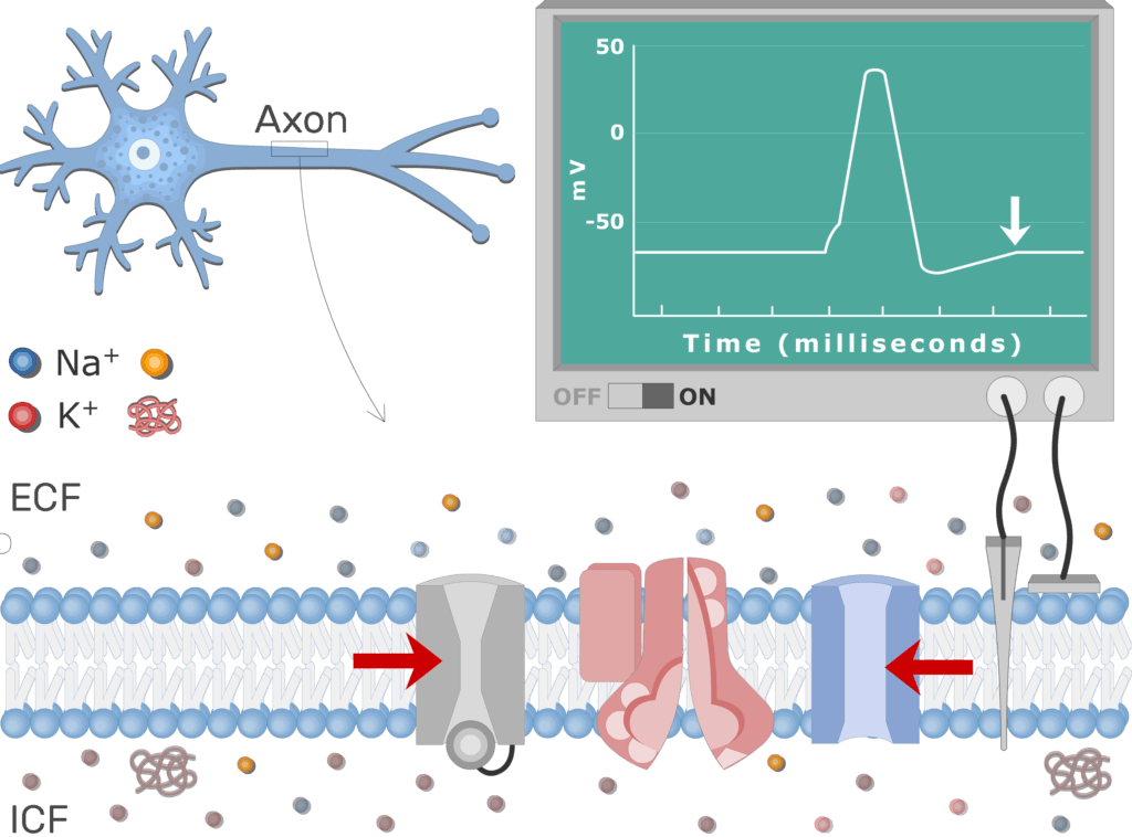
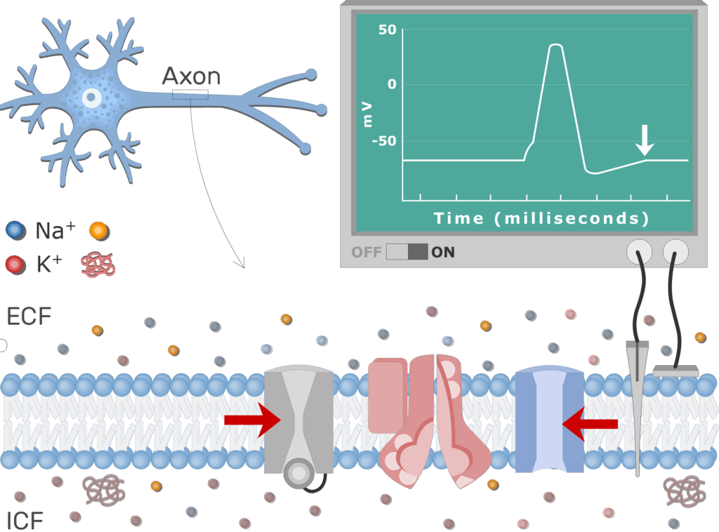
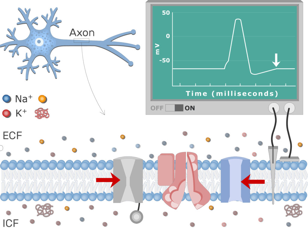
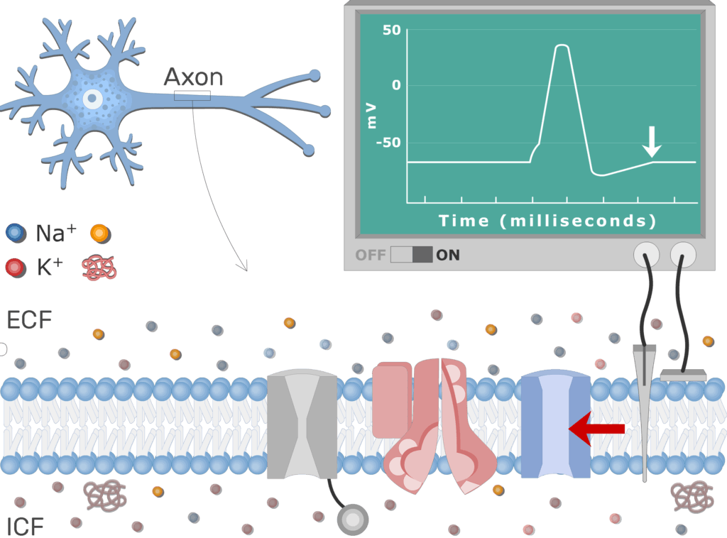
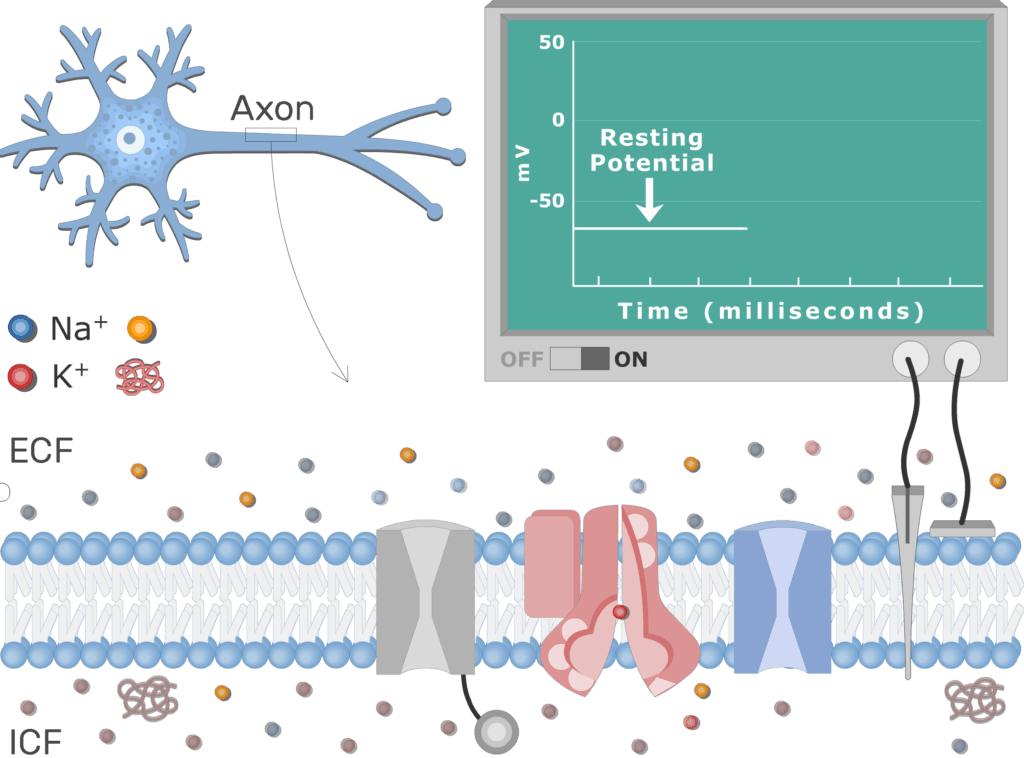
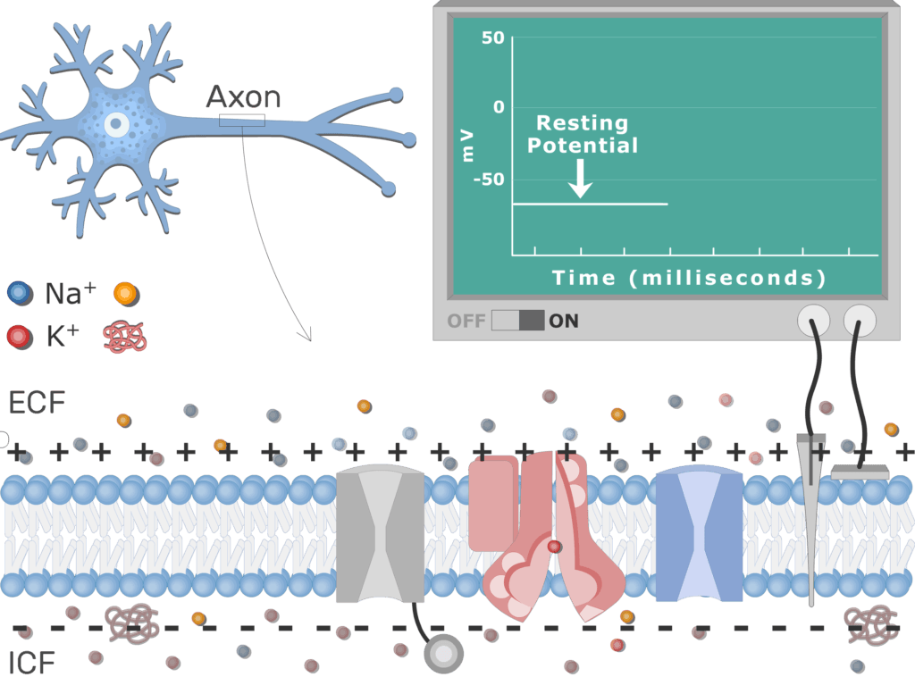
At rest, the axon membrane is slightly ![]() polarized to about -70mV,
polarized to about -70mV, ![]() meaning the intracellular fluid (ICF) is relatively negative to the extracellular fluid (ECF).
meaning the intracellular fluid (ICF) is relatively negative to the extracellular fluid (ECF).
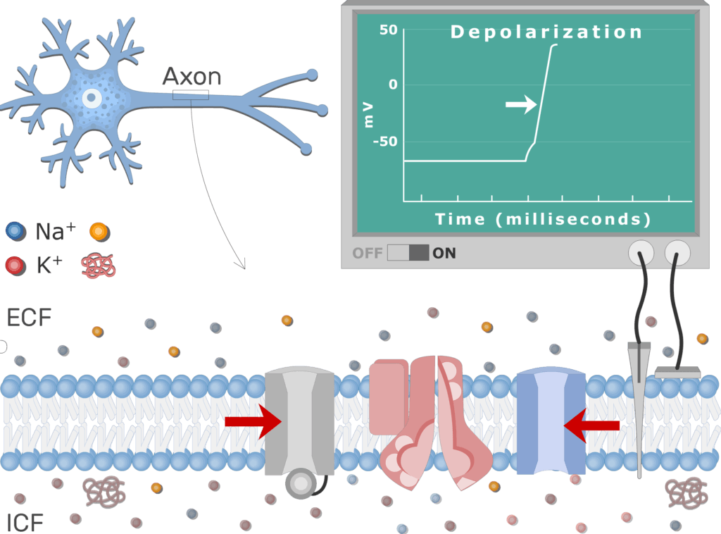
- An action potential occurs when a portion of the membrane rapidly depolarizes and then repolarizes again to the original resting state.
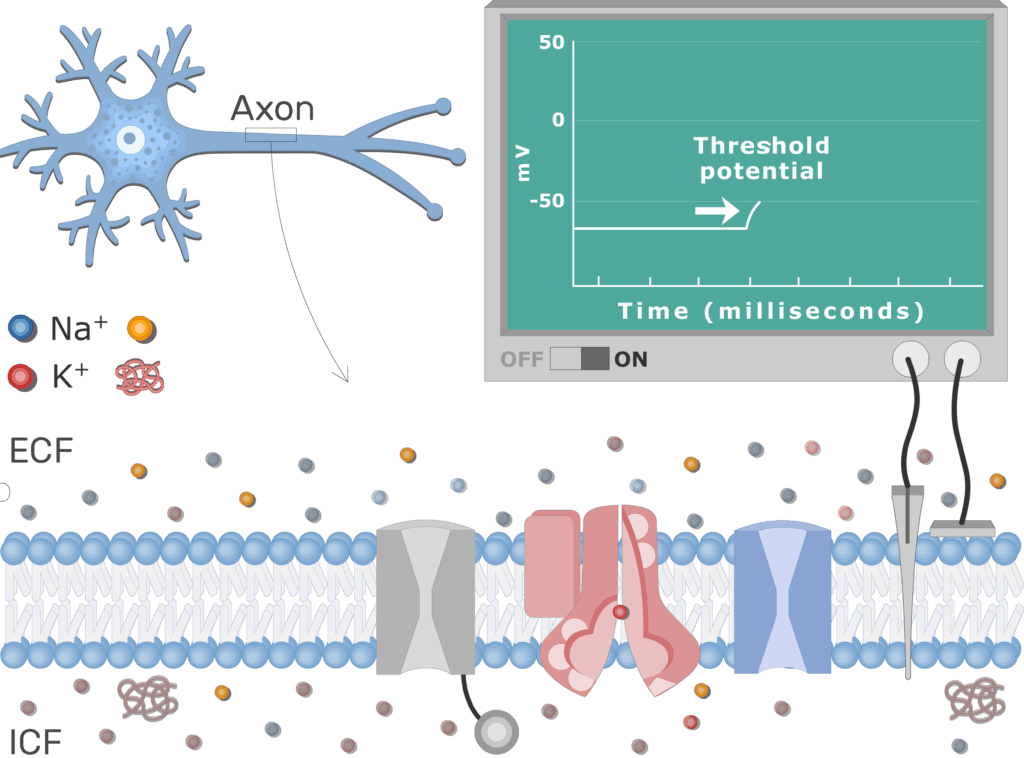
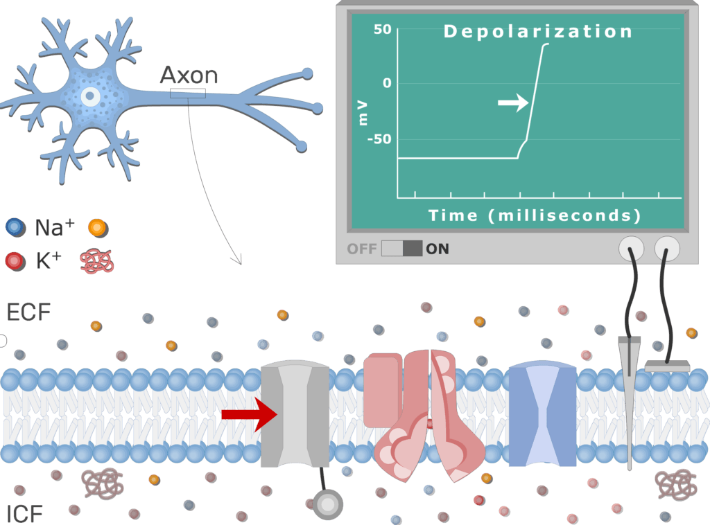
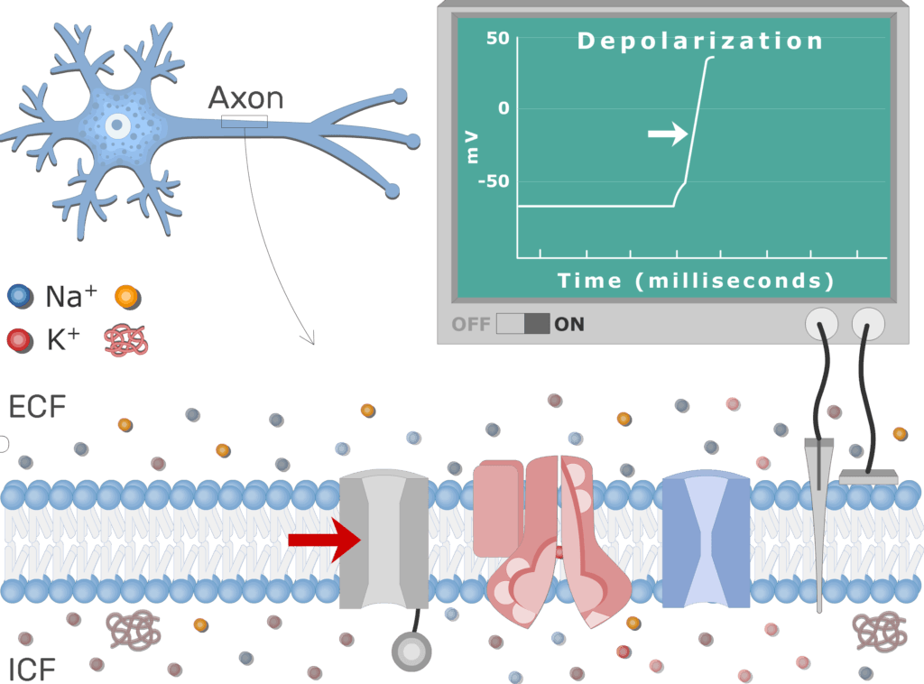
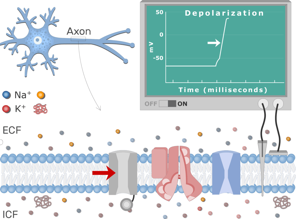
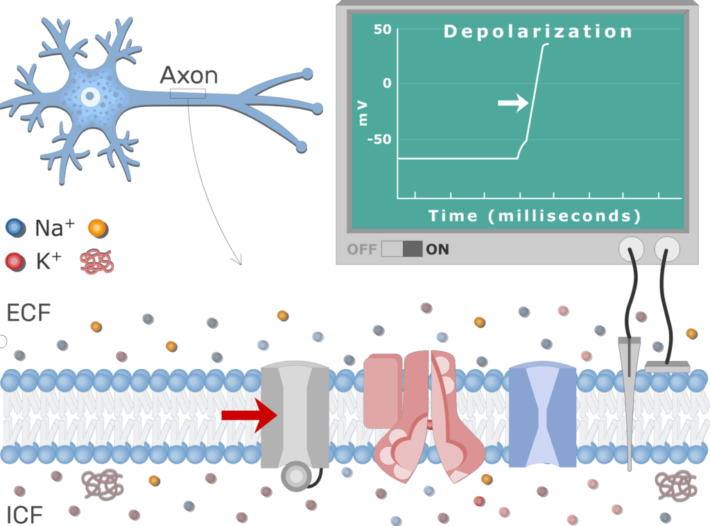
- The process is initiated by a threshold level
 stimulus, such as a nearby change in membrane potential (threshold potential, local potential).


- At threshold (about -55mV), many
 Na+ voltage-gated channels open.
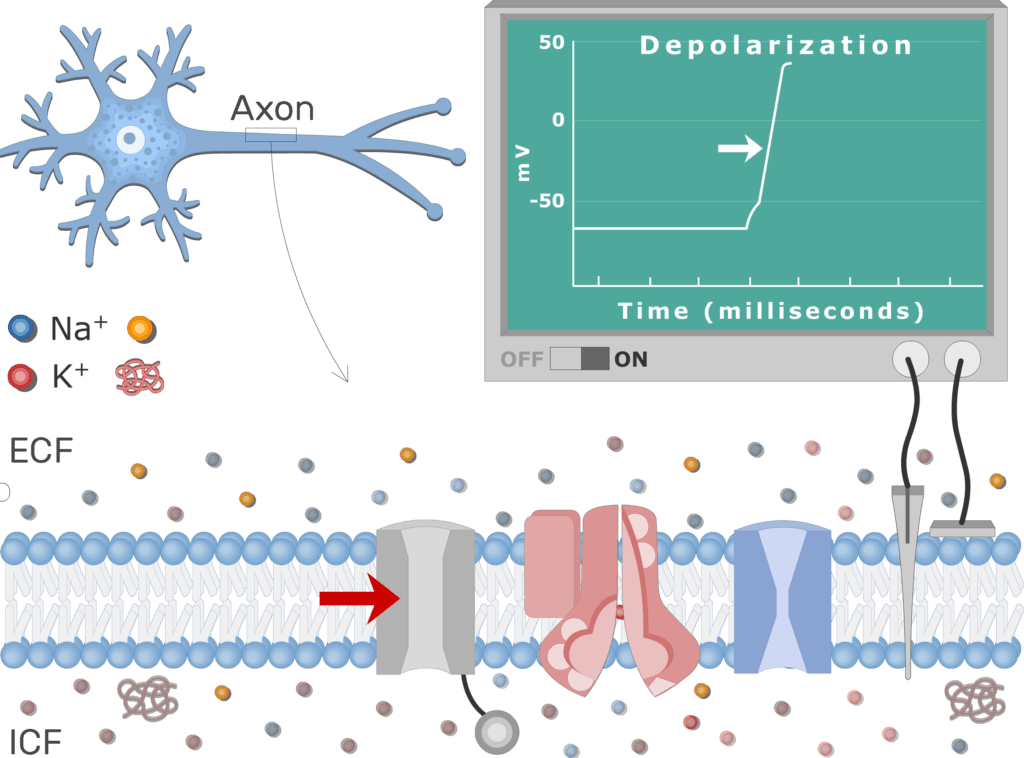
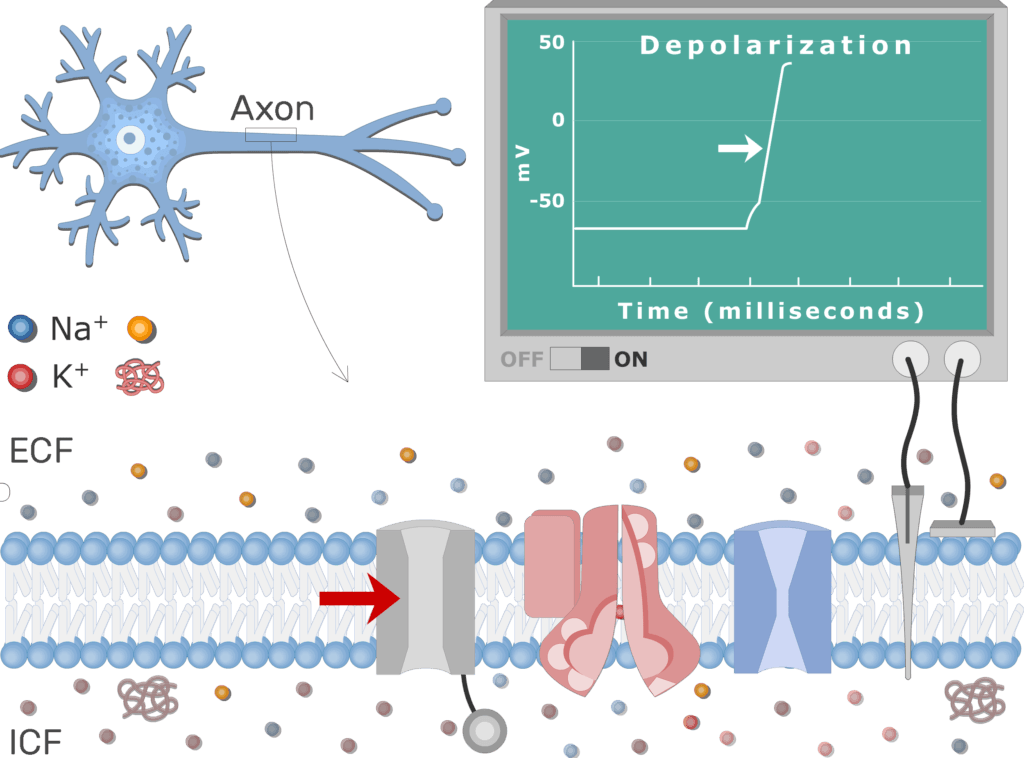
- Na+ ions entering the cell make the membrane potential less negative. More Na+ channels open as result and a cycle of depolarization develops.
Discover the anatomy of the nervous system with these interactive quizzes and labeled diagrams.
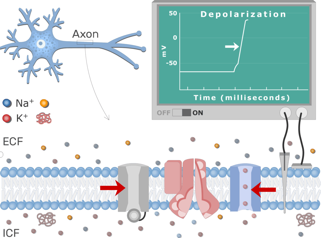
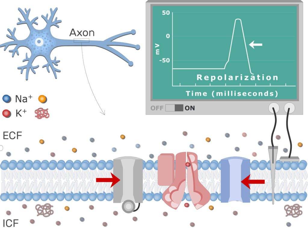
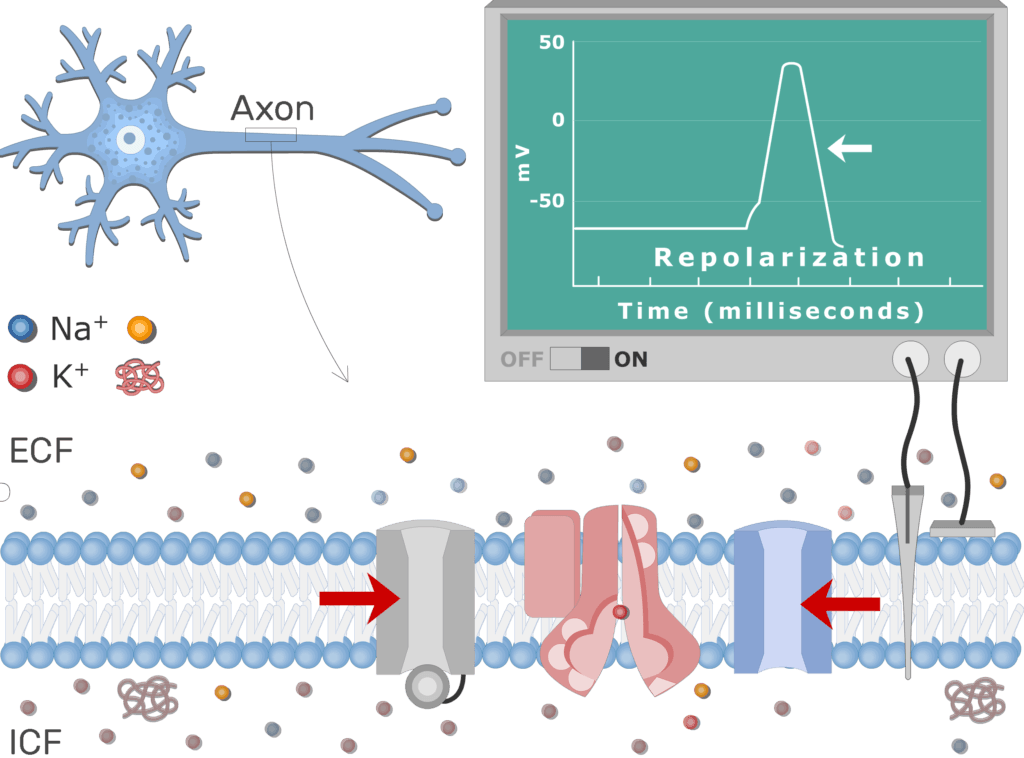
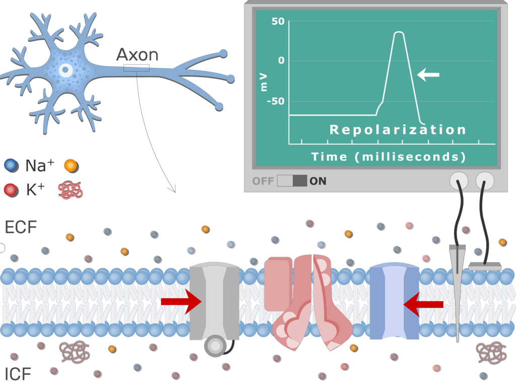
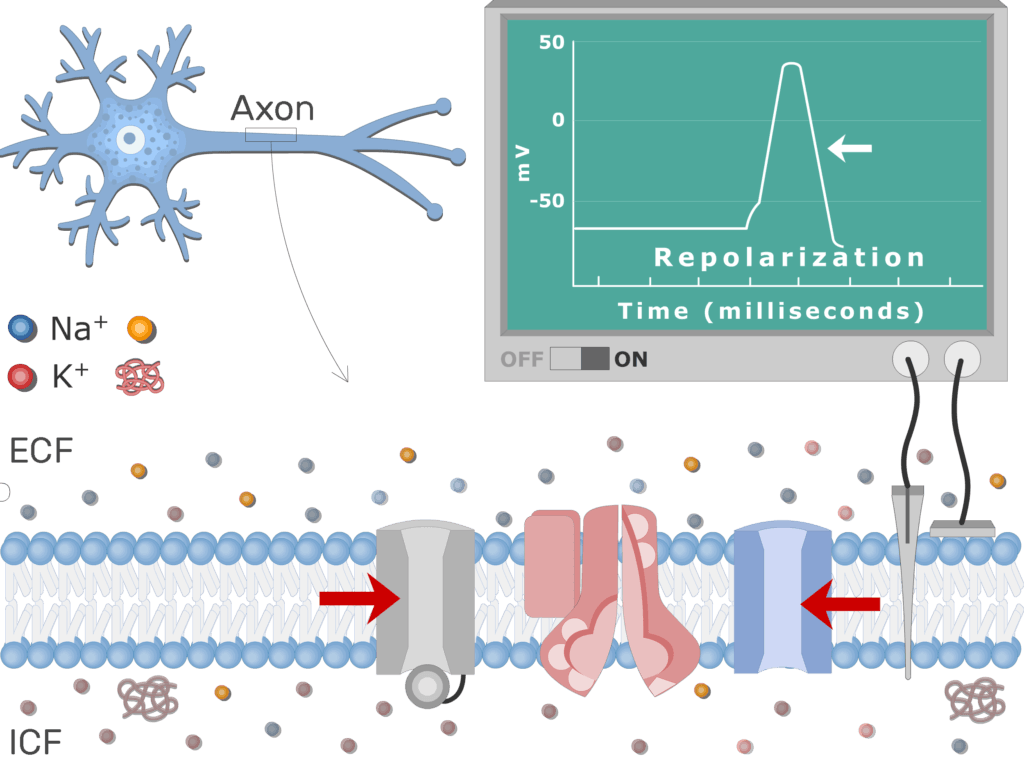
- When the membrane potential reaches about +30mV (reverse polarization), the timed Na+ channels close due to inactivation and the Na+ influx stops.
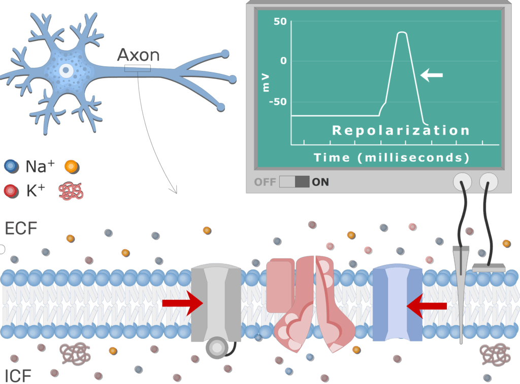
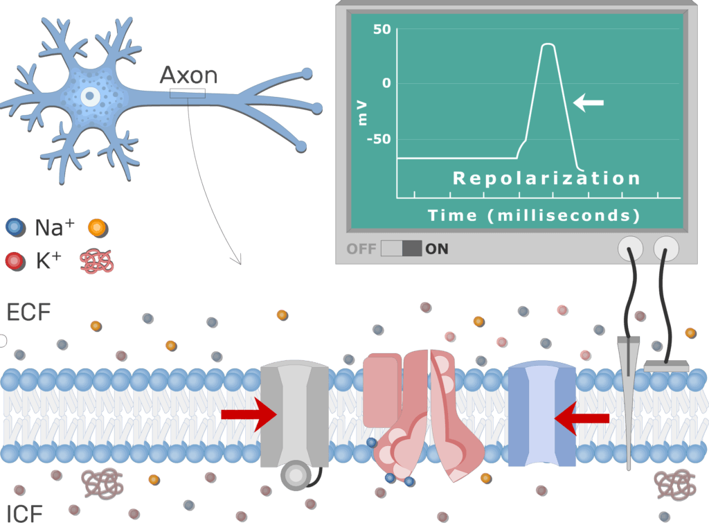
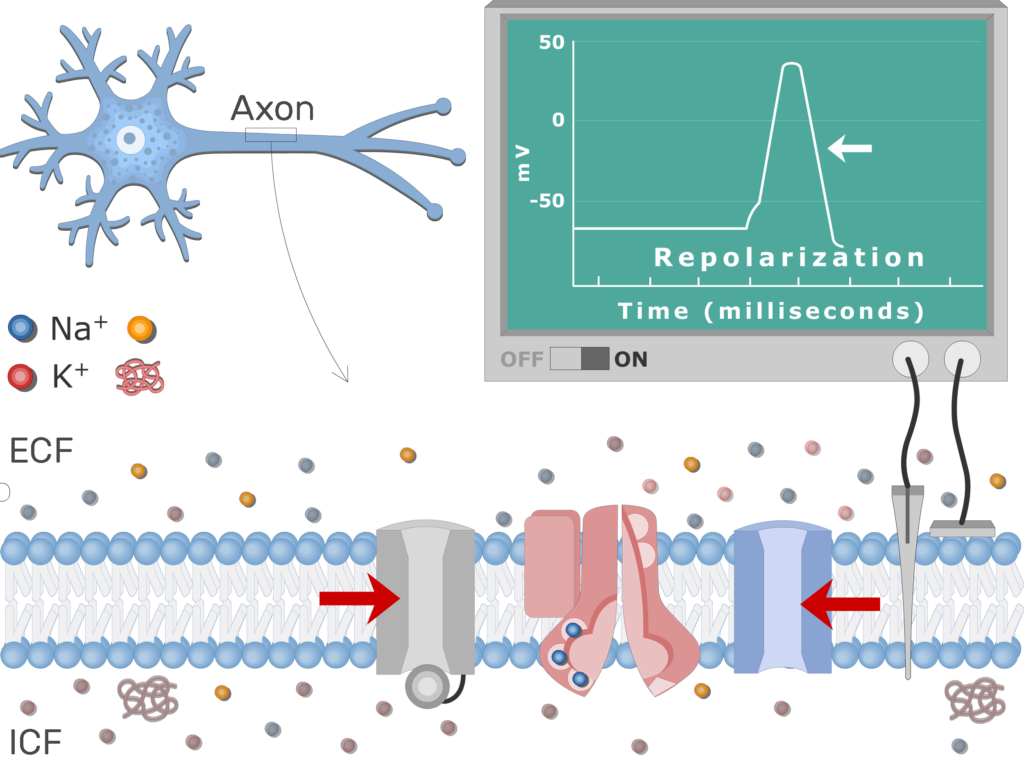
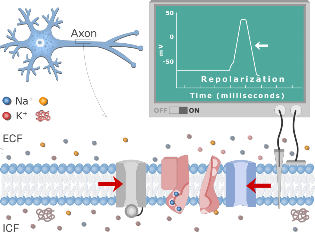
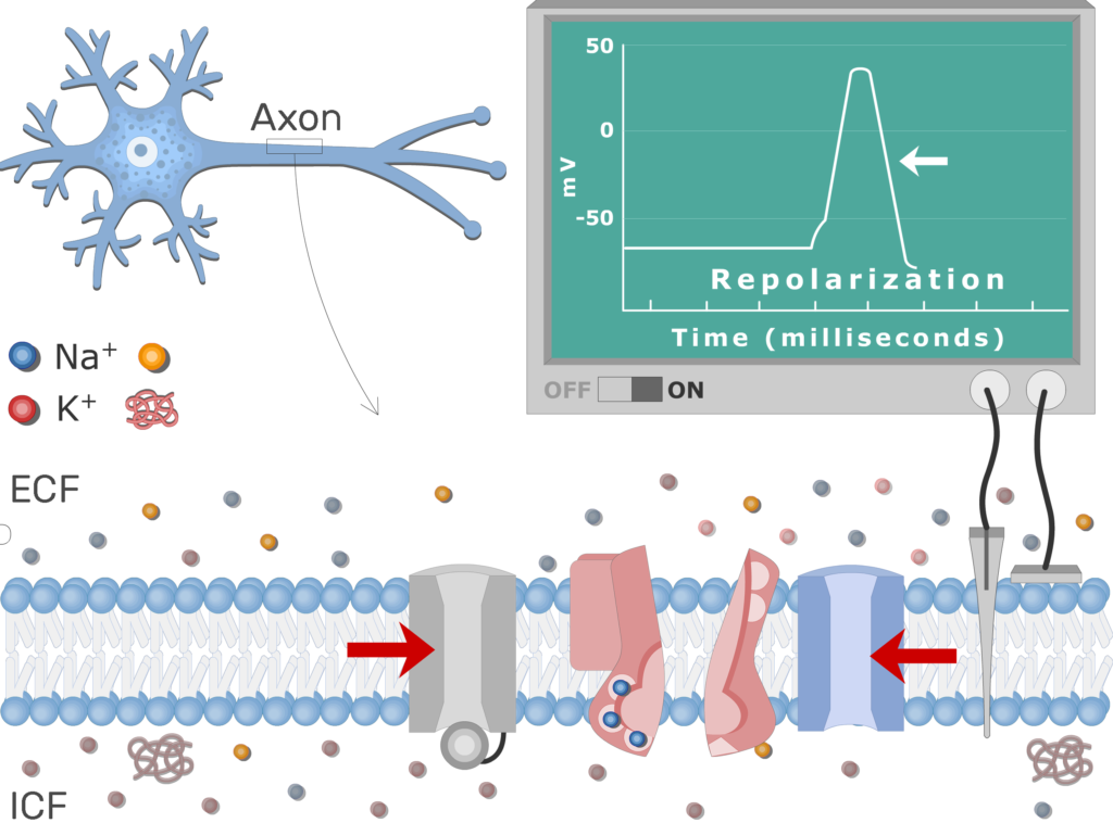
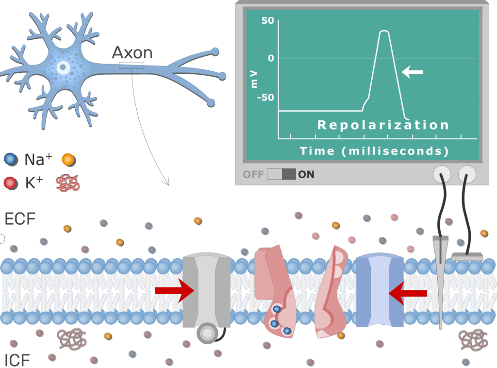
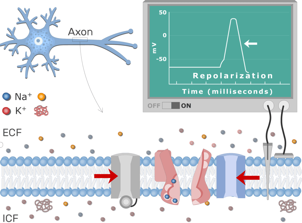
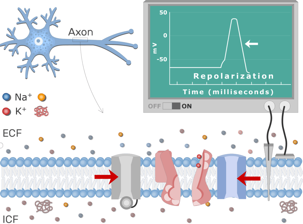
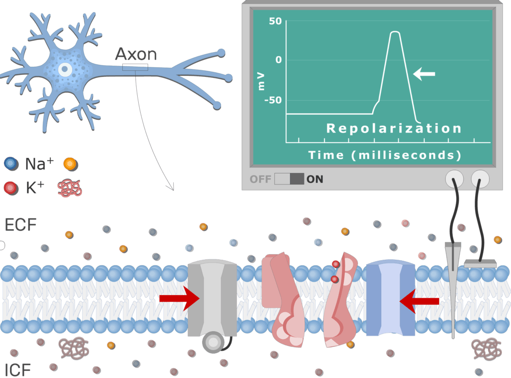
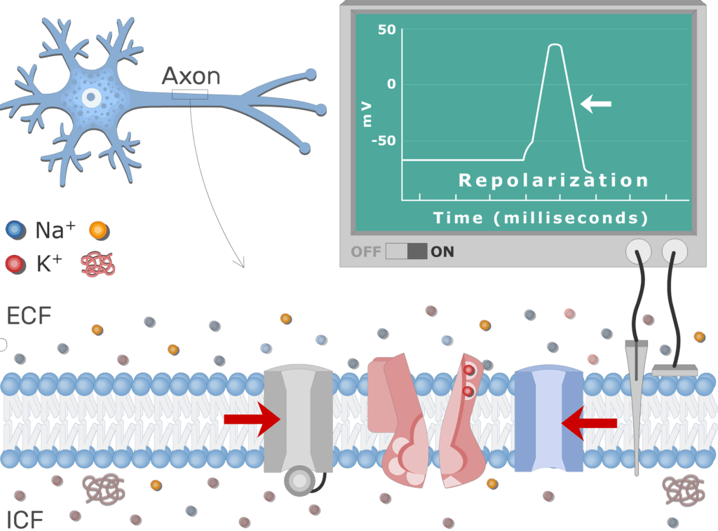
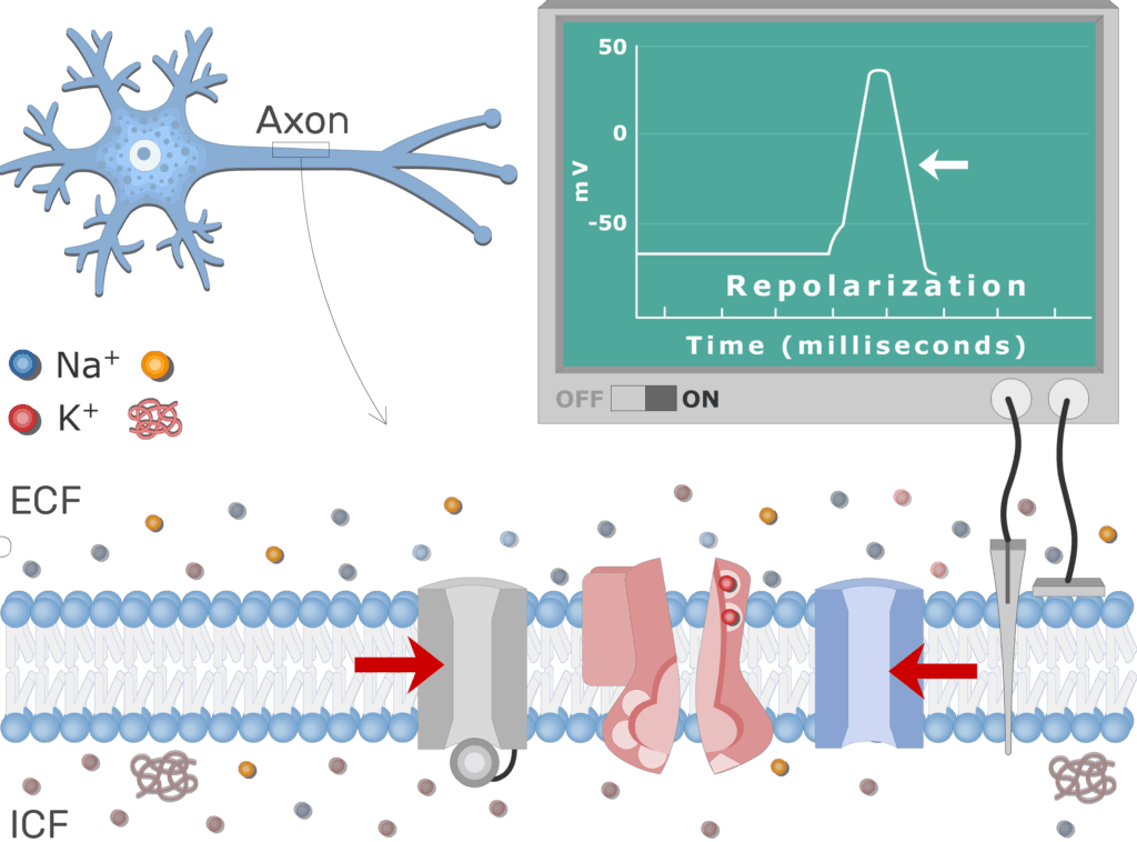
- K+ voltage-gated channels open as Na+ channels close, in a delayed response to the original stimulus.
- K+ ions diffuse out of the cell and the efflux of positive charges causes the membrane to rapidly repolarize.

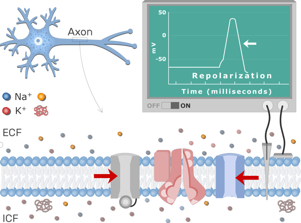
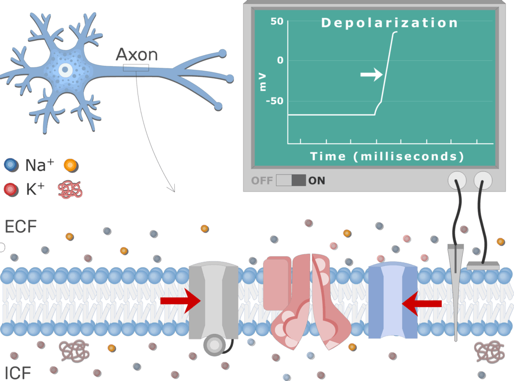
- The K+ channels are slow to close, so the membrane briefly hyperpolarizes.
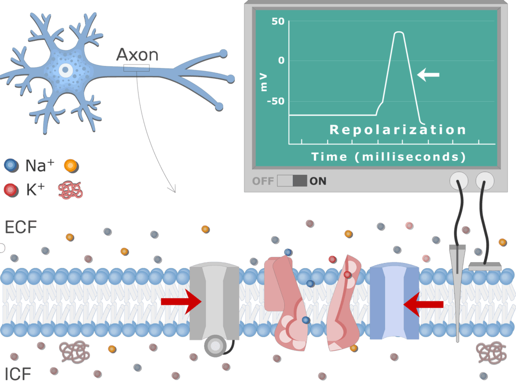
- As the K+ channels close, Na+/K+ pumps actively transport Na+ ions out of the cell and K+ ions into the cell.
- The ion exchange helps re-establish the ion diffusion gradients and resting membrane potential.
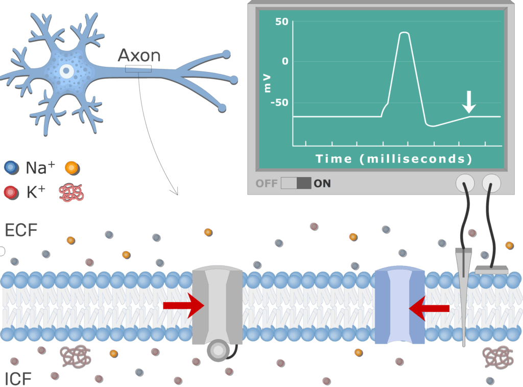
- Lastly, the Na+ channels close (dein-activate) as the resting potential returns.
Related Articles
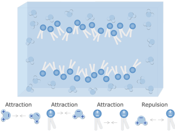
Water-Cell Membrane Interactions
Water-Cell Membrane Interactions; explained beautifully in an illustrated and interactive way.
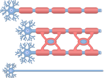
Unmyelinated and Myelinated Axons
Unmyelinated and Myelinated Axons; explained beautifully in an illustrated and interactive way.
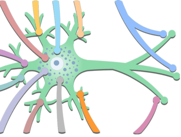
Types of Synaptic Contacts
Types of Synaptic Contacts; explained beautifully in an illustrated and interactive way.