


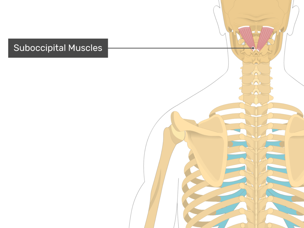
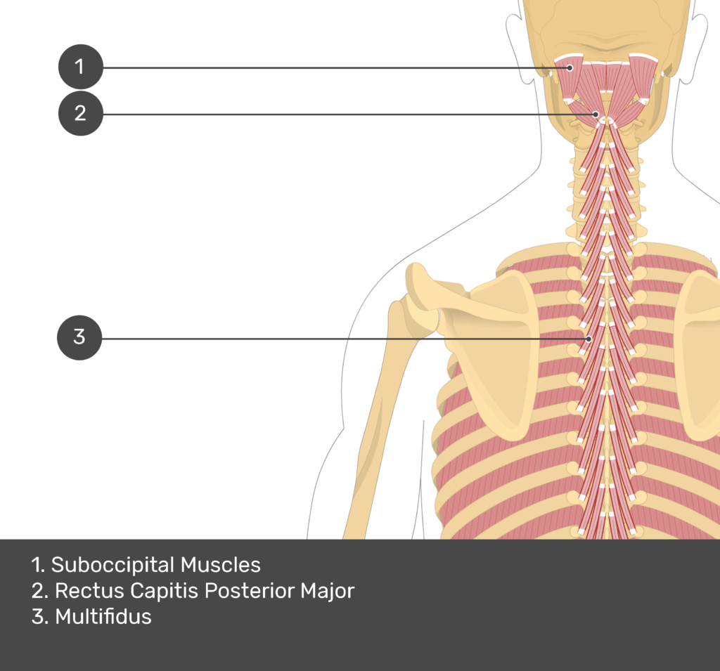
Obliquus Capitis Superior Muscle
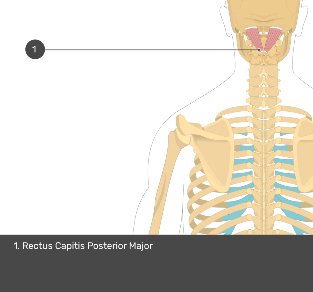
Rectus Capitis Posterior Major
Last update:
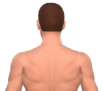
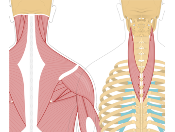
The rectus capitis posterior major is a broad, flat muscle situated in the back of the neck.
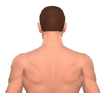
 Rectus Capitis Posterior Major

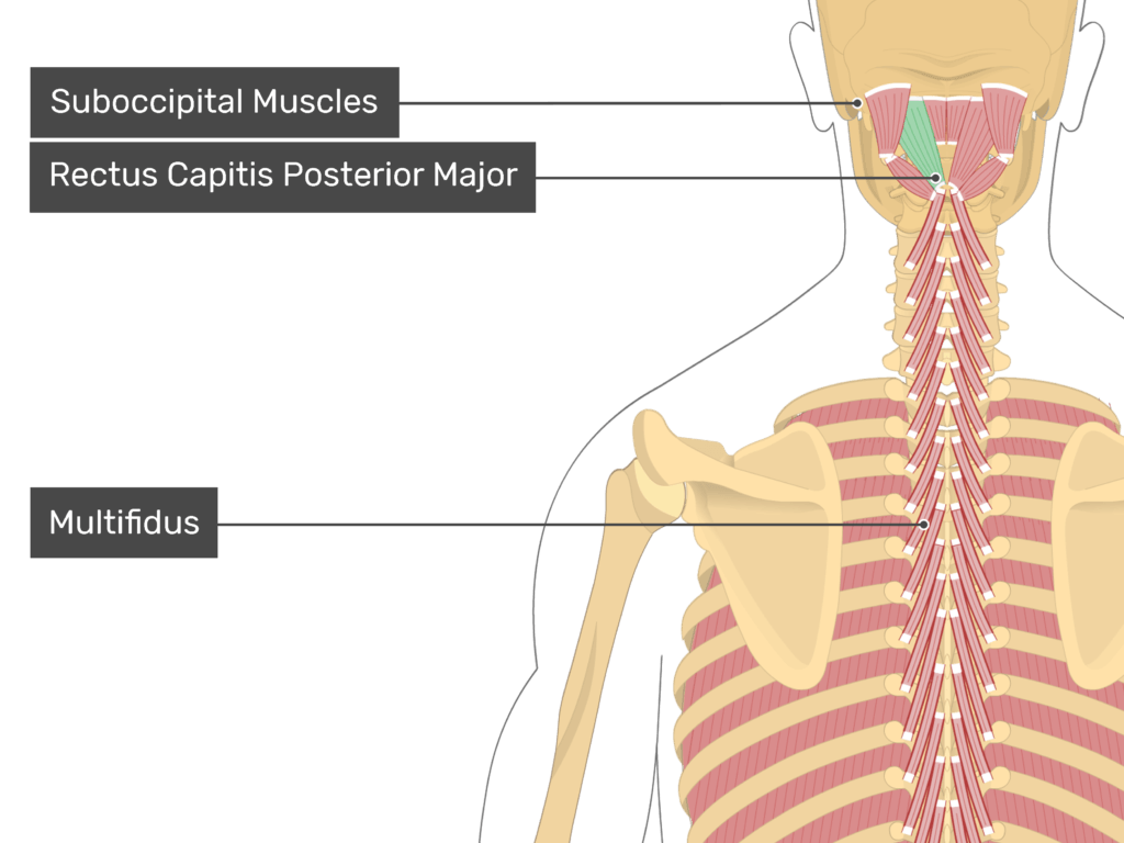
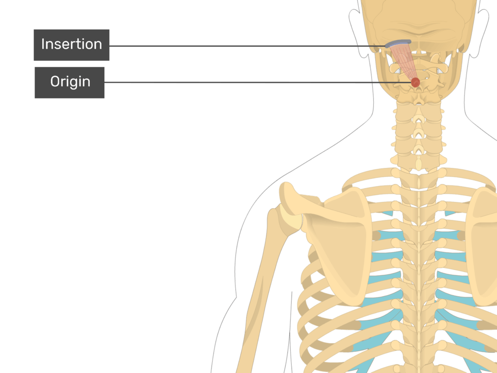
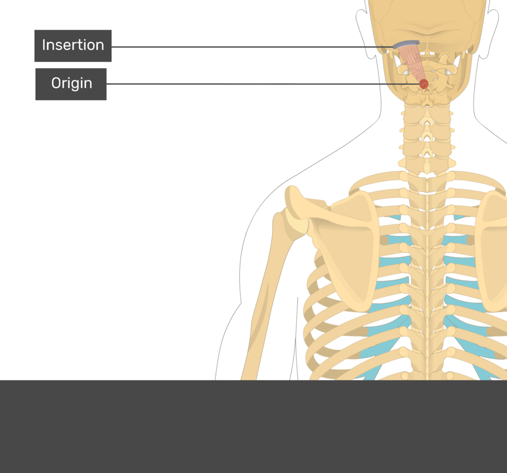
Attachments of the Rectus Capitis Posterior Major Muscle
Origin
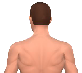
a. Spinous process of the axis vertebra (C2).
Insertion
a. Lateral portion of inferior nuchal line of occipital bone.
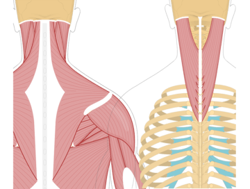
Actions of the Rectus Capitis Posterior Major:
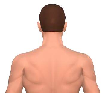
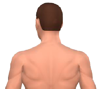
a. Bilaterally ![]() extends the head.
extends the head.
Learning muscle anatomy is hard – but it can be made easier! Find out how to save time with these muscle anatomy reference charts.
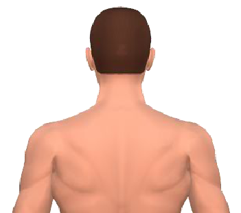
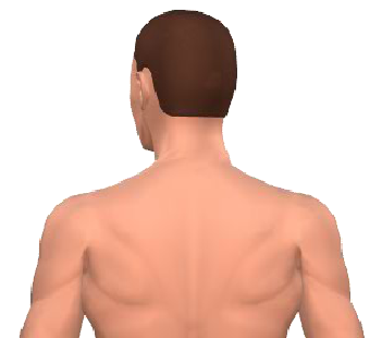
b. Unilaterally ![]() rotates head to same side (ipsilateral rotation).
rotates head to same side (ipsilateral rotation).
Nerve to the Rectus Capitis Posterior Major Mucle:
a. Suboccipital nerve or posterior (dorsal) primary ramus of C1.
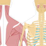
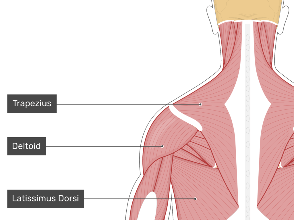
An Overview of the Muscles of the Neck


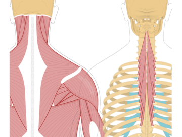
Test yourself while observing the Rectus Capitis Posterior Major muscle
Related Articles
Splenius Cervicis Muscle
A tutorial on the position, actions, attachments and innervation of the Splenius Cervicis Muscle with the aid of detailed anatomical illustrations.
Splenius Capitis Muscle
A tutorial on the position, actions, attachments and innervation of the Splenius Capitis with the aid of detailed anatomical illustrations.
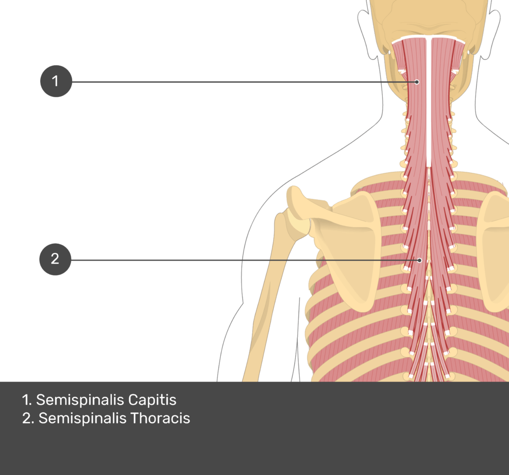
Semispinalis Thoracis Muscle
A tutorial on the position, actions, attachments and innervation of the Semispinalis Thoracis with the aid of detailed anatomical illustrations.
