


Primary Somatosensory Cortex
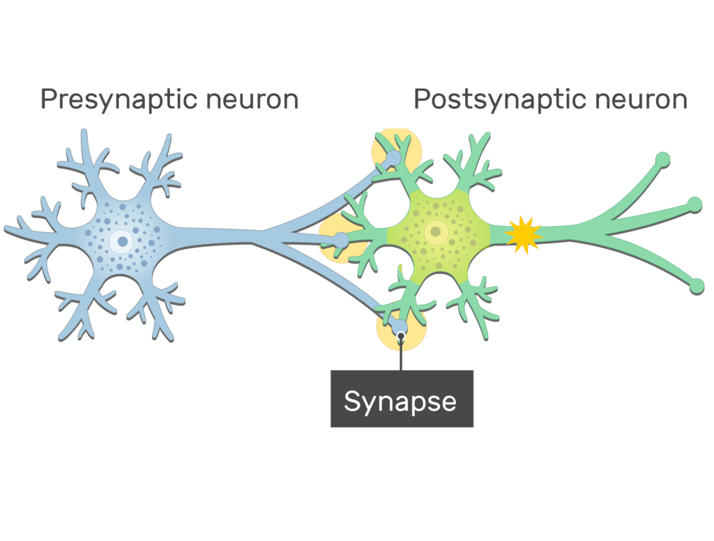
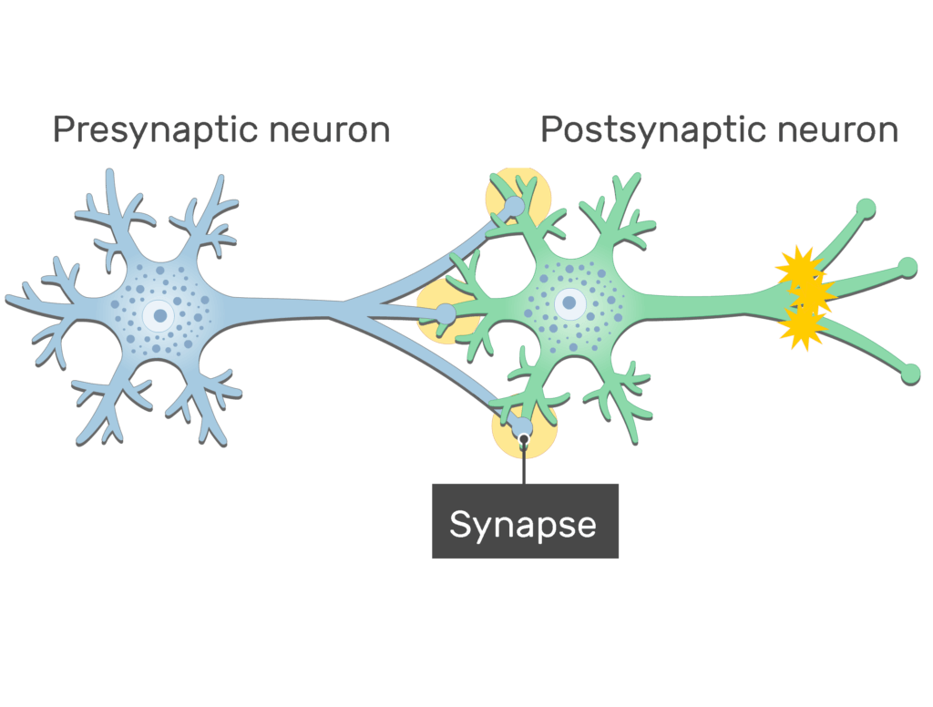
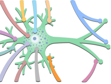
General Structure of a Neuron Synapse
Last update:
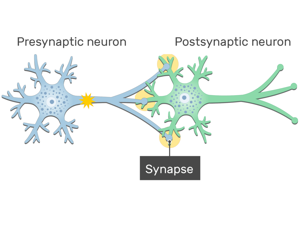
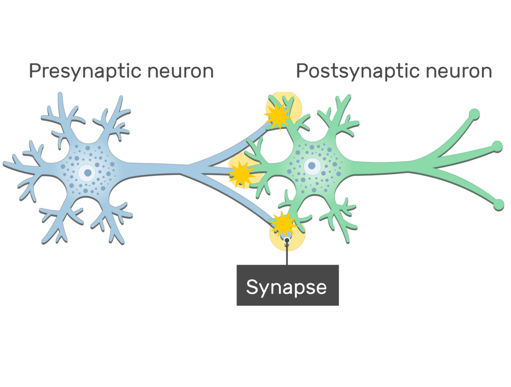
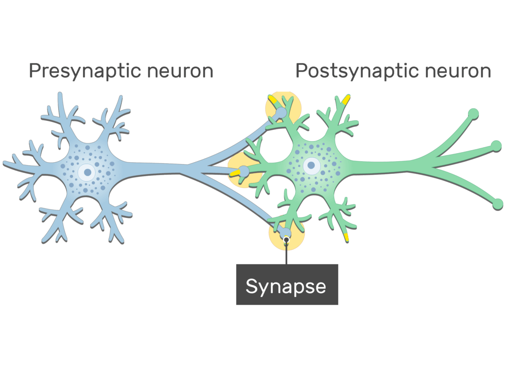
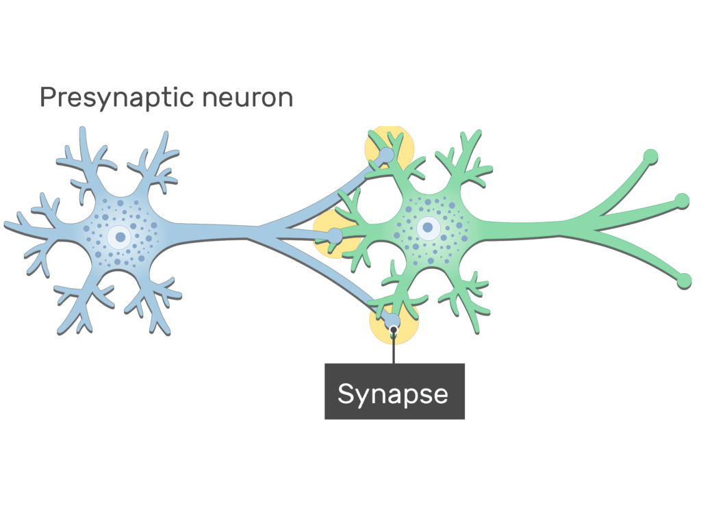
A ![]() synapse is a junction formed between two neurons, which allows the cells to communicate.
synapse is a junction formed between two neurons, which allows the cells to communicate.
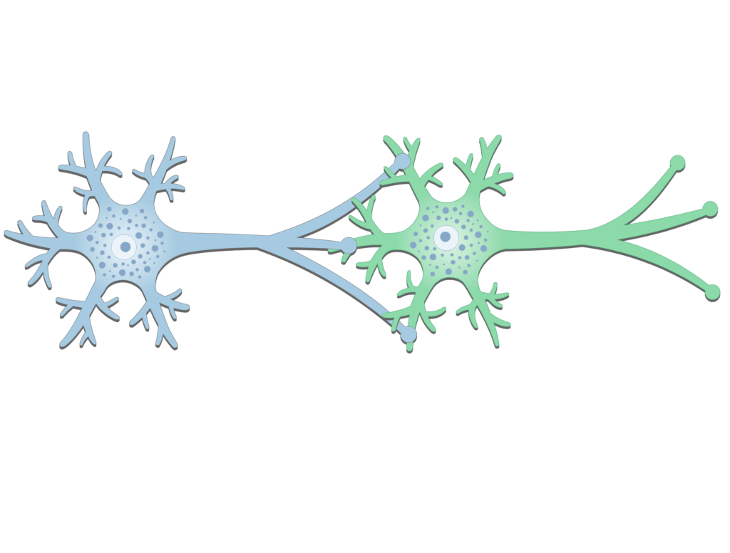

- The neuron approaching the synapse is called the
 presynaptic neuron.
- On the other side of the synapse is the postsynaptic neuron.
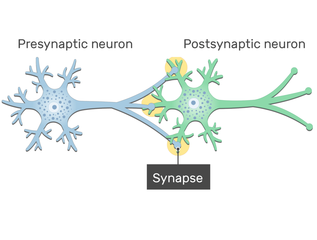
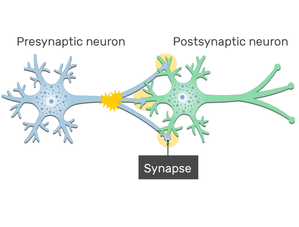
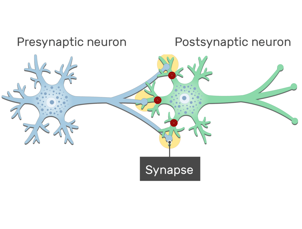
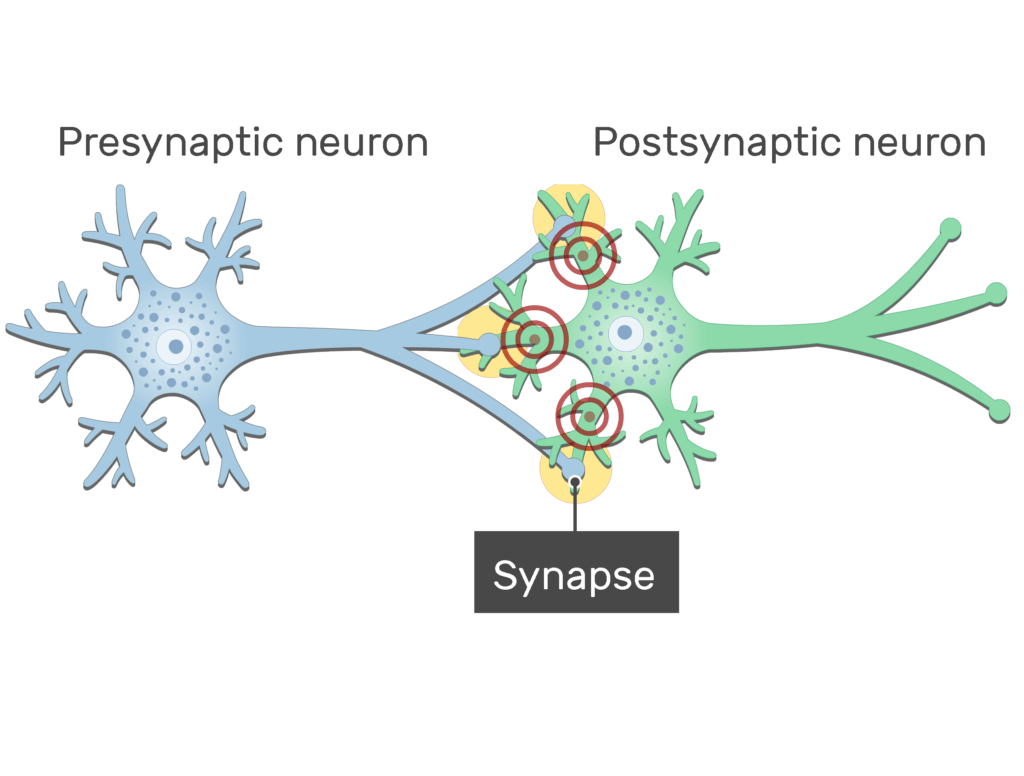
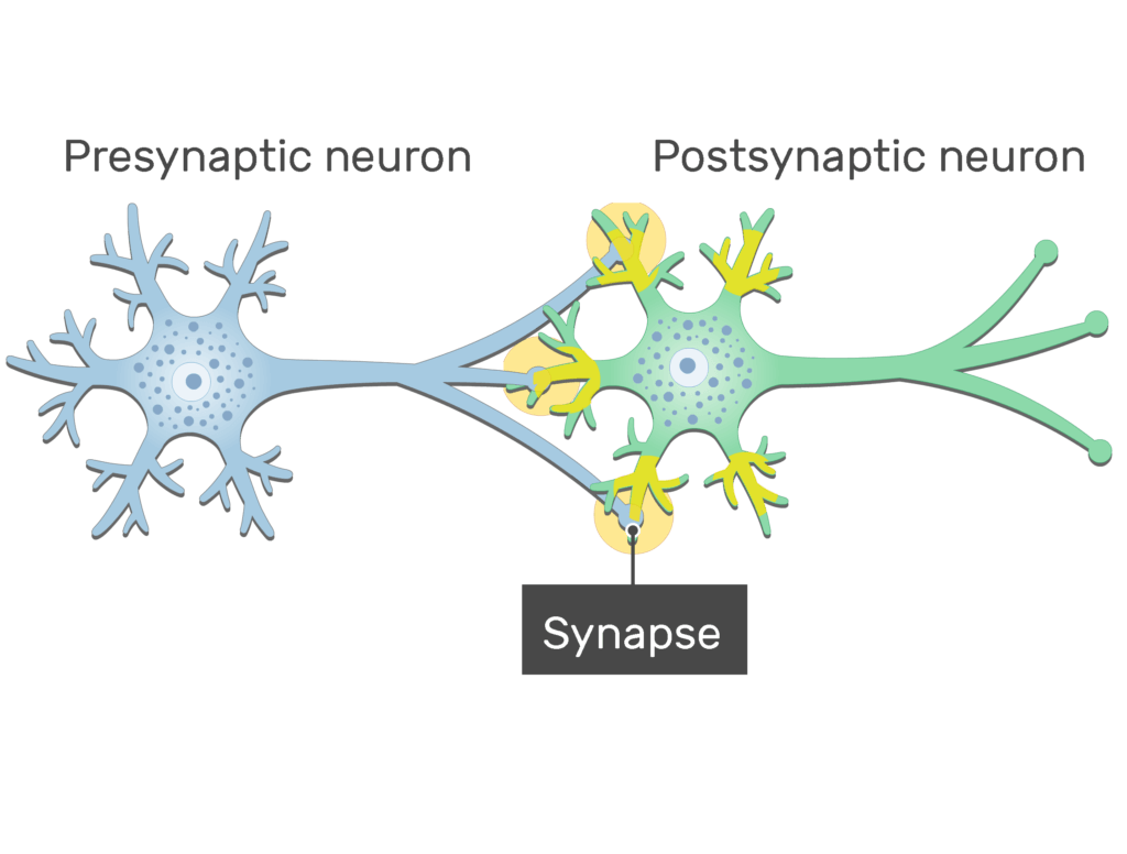
- At the synapse, the presynaptic neuron
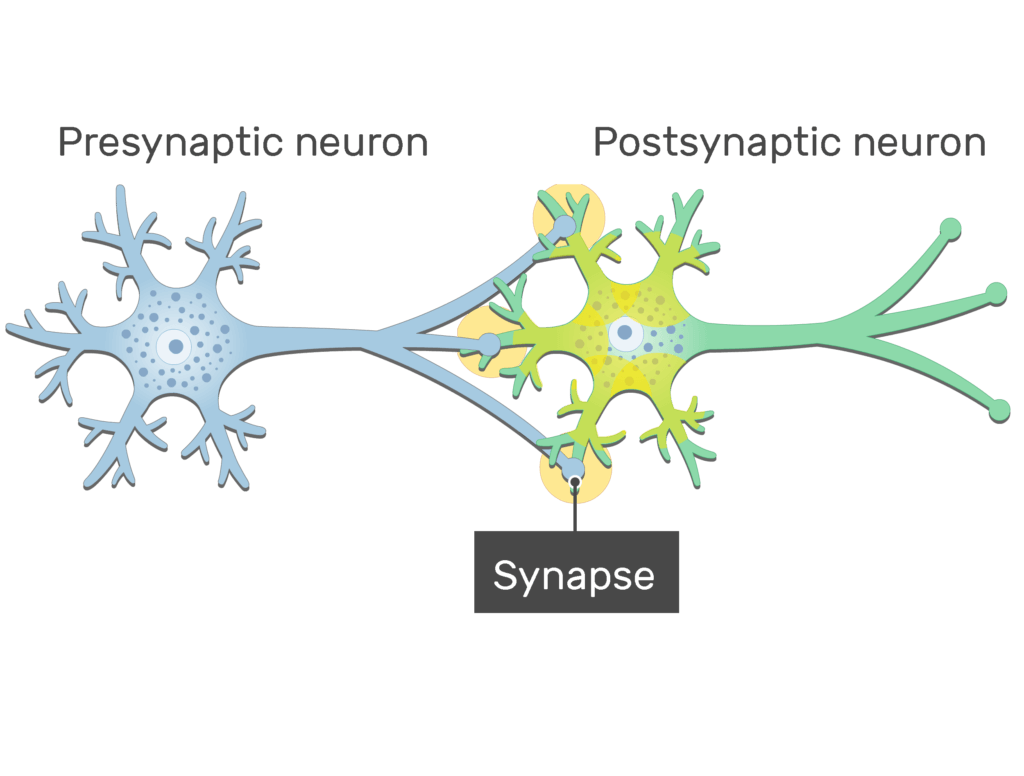
 sends (or transmits) chemical or electrical signals to the postsynaptic neuron.
Master nervous system anatomy in half the time with these interactive quizzes, diagrams and worksheets.
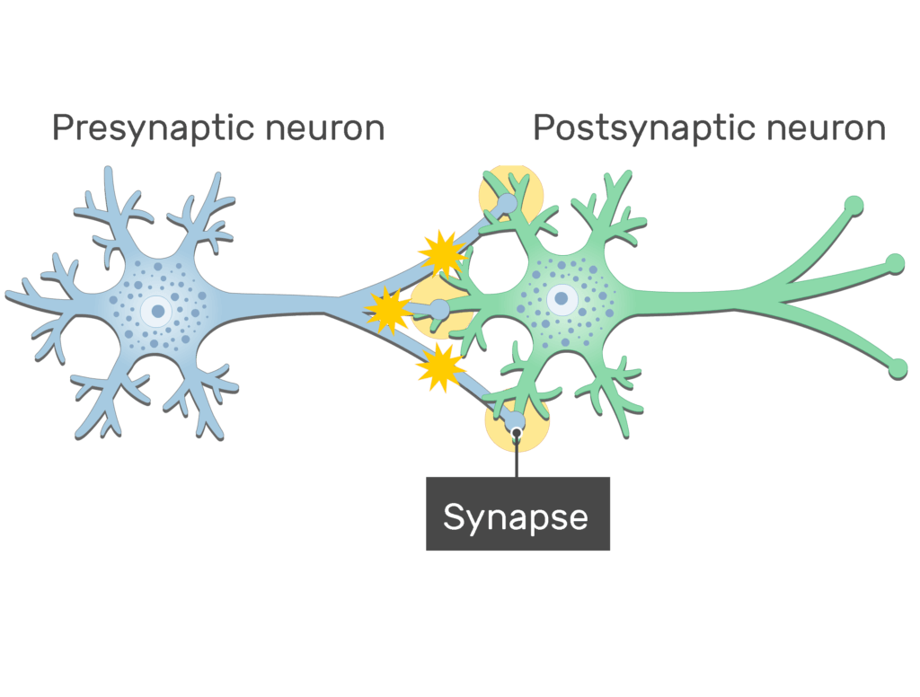
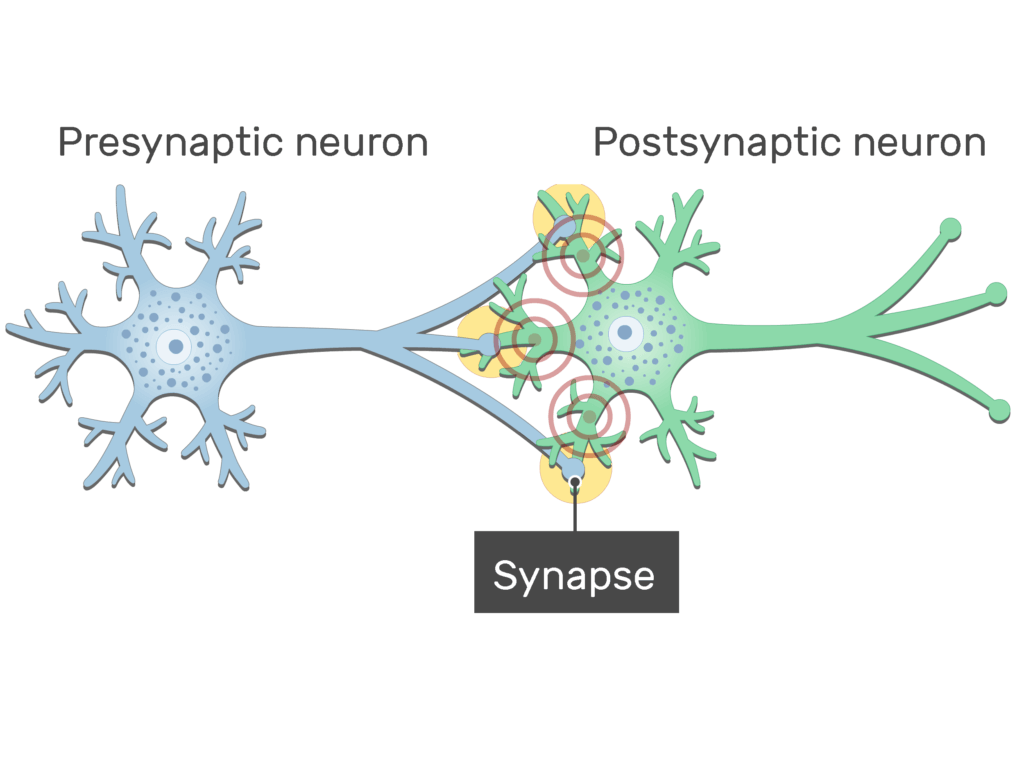
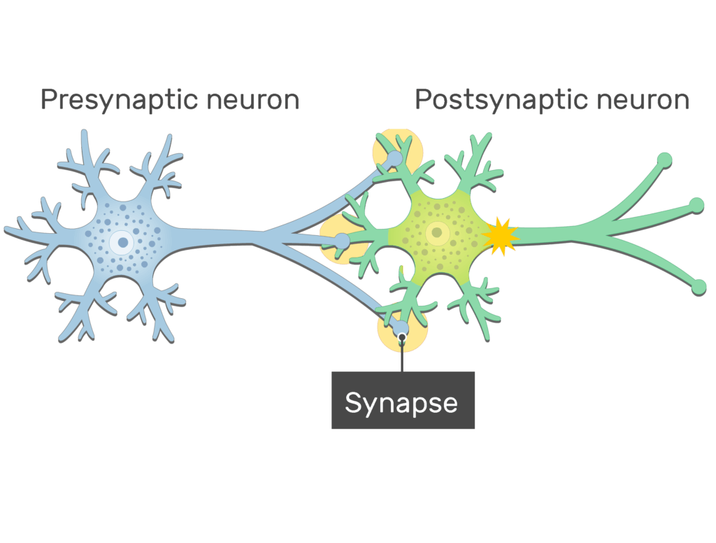
- The signals received by the postsynaptic neuron produce either an inhibitory or excititory repsonse.
Related Articles
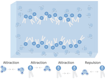
Water-Cell Membrane Interactions
Water-Cell Membrane Interactions; explained beautifully in an illustrated and interactive way.
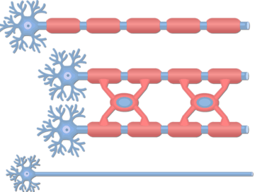
Unmyelinated and Myelinated Axons
Unmyelinated and Myelinated Axons; explained beautifully in an illustrated and interactive way.
Types of Synaptic Contacts
Types of Synaptic Contacts; explained beautifully in an illustrated and interactive way.