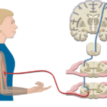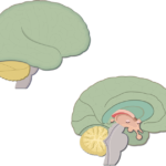
Major brain parts and their functions
Spinal Cord Gray Matter Anatomy & Functions
Last update:
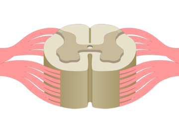
The gray matter of the spinal cord is a vital part of the central nervous system and is involved in muscle movement, sensory information like fine touch, proprioception and vibration, and more.
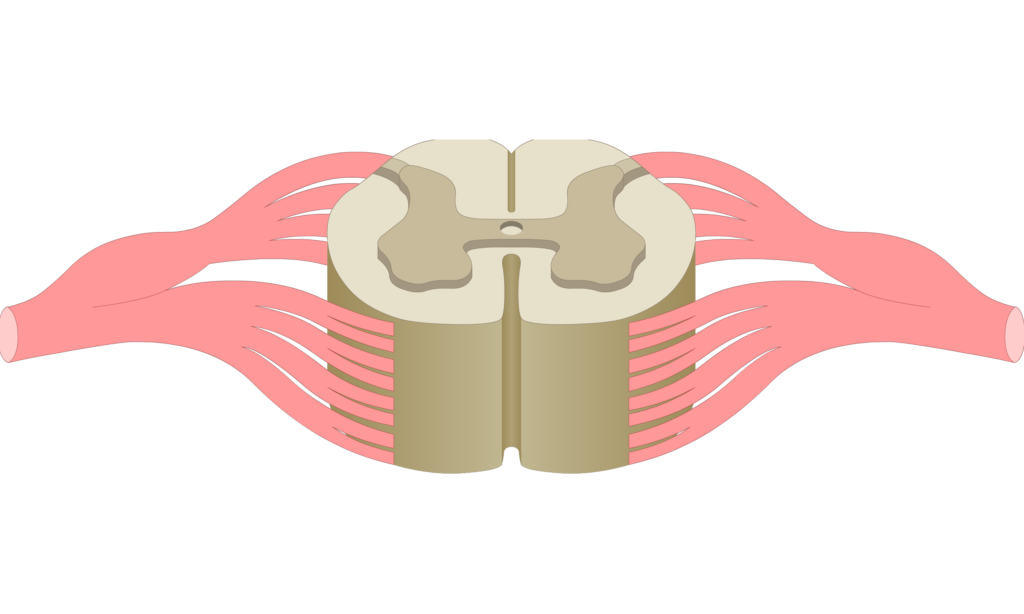
Anatomy of the spinal cord gray matter:
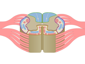
- Each arm (or extension) of the gray matter in the spinal cord is referred to as a horn.
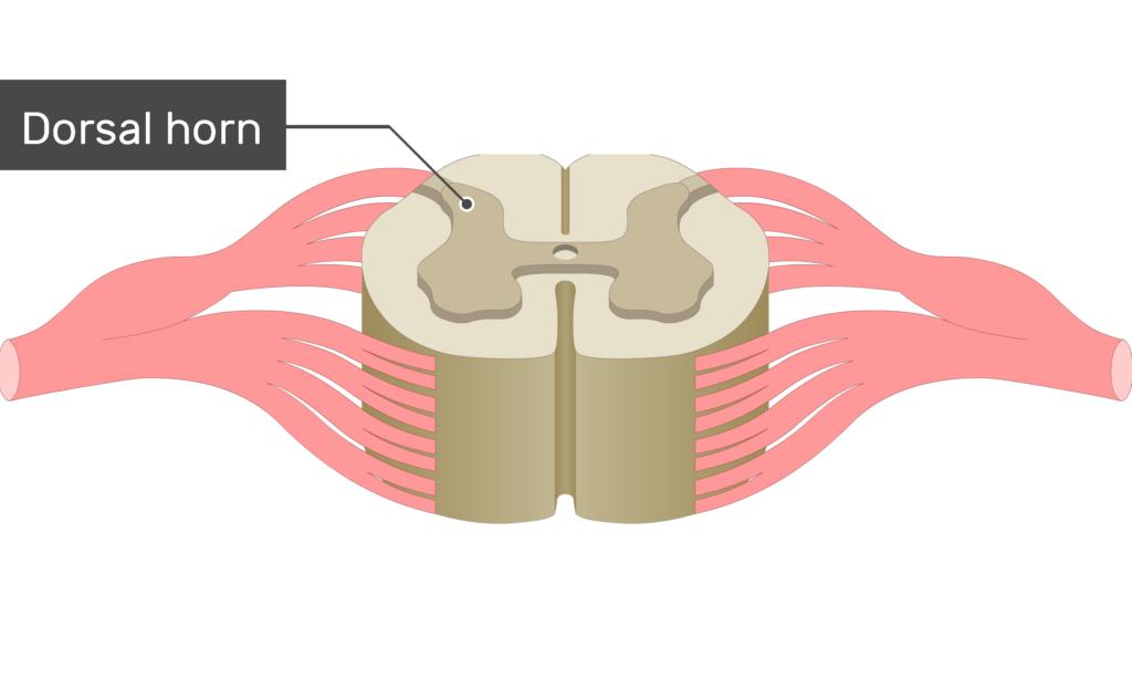
- Projecting towards the back of the spinal cord are the
 dorsal horns (or posterior horns).
 - Projecting towards the front are the ventral horns (or anterior horns).
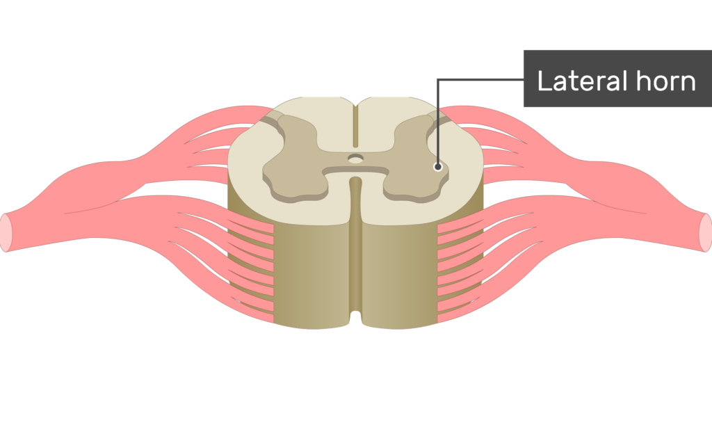
- In the thoracic and upper lumbar regions of the cord, an additional pair of side projections occur, which are called the lateral horns.
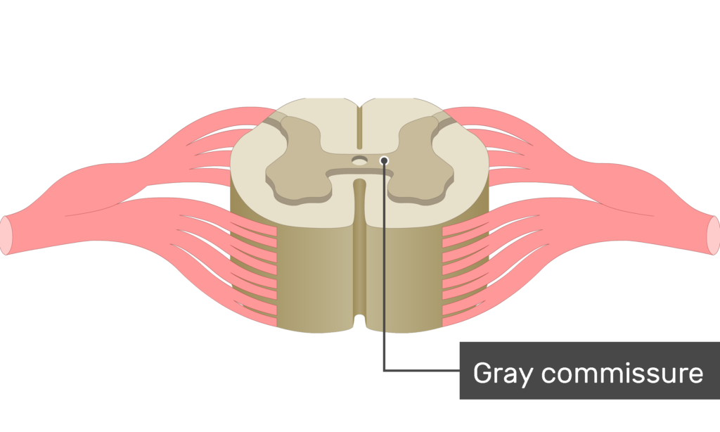
- A narrow band of gray matter known as the gray commissure stretches across of the center of the spinal cord and connects the two sets of horns.
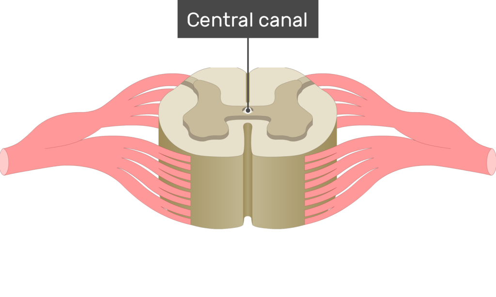
- In the middle of the gray commissure is the central canal, which contains cerebral spinal fluid.
Understand white and grey matter with these interactive spinal cord quizzes, diagrams and worksheets.

Functions of the spinal cord gray matter:
- The gray matter is the area of the spinal cord where many types of neurons synapse.
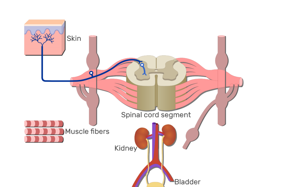
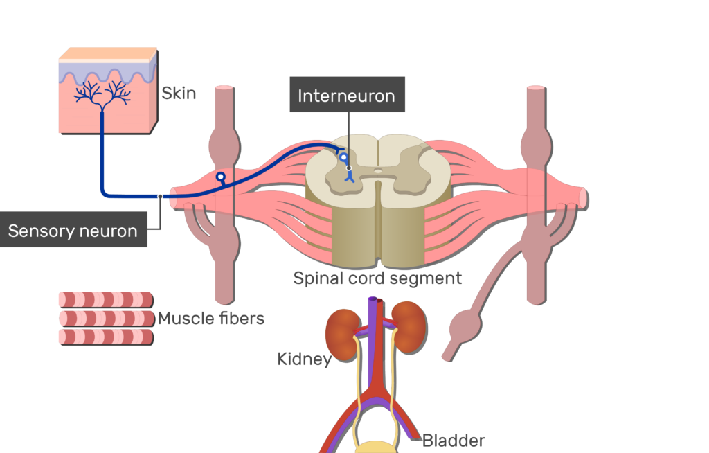
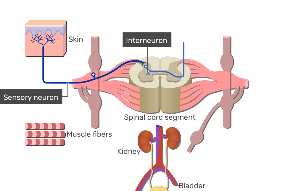
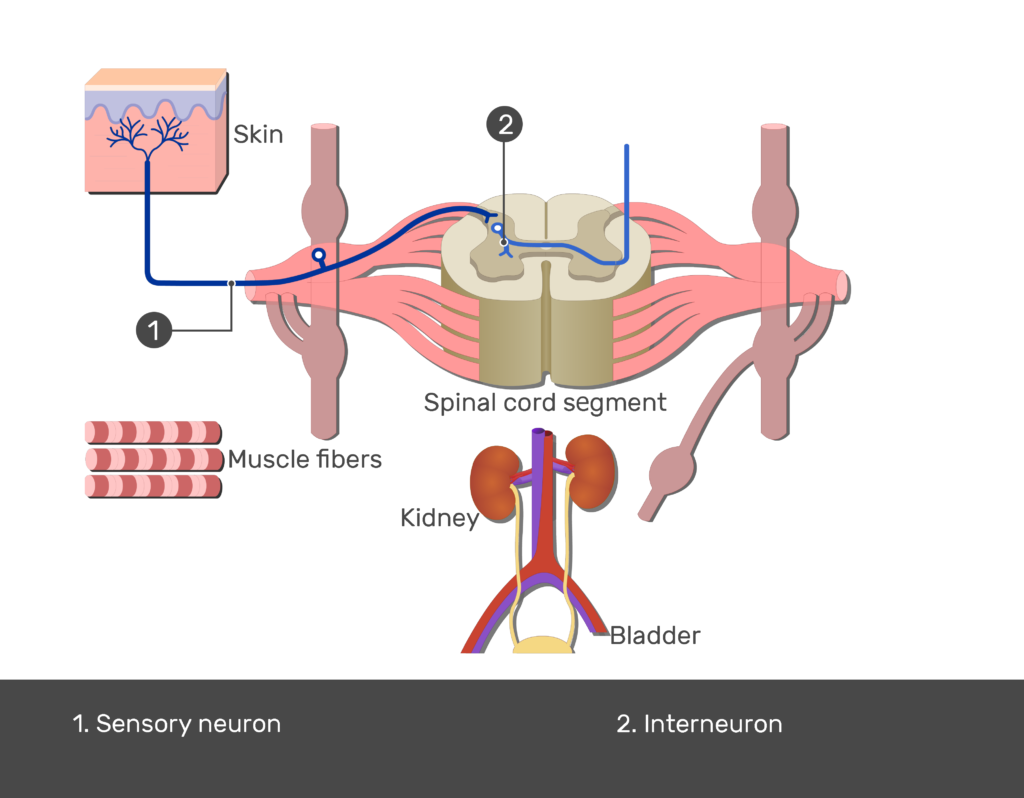
- In the dorsal horns (or posterior horns), many incoming sensory neurons synapse with interneurons, which then distribute information to other parts of the spinal cord and brain.
- Some of the interneurons use the gray commissure to cross over to the other side of the spinal cord.
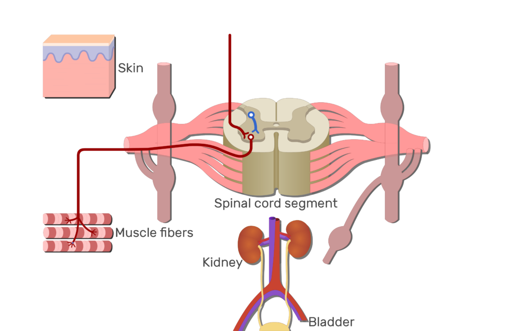
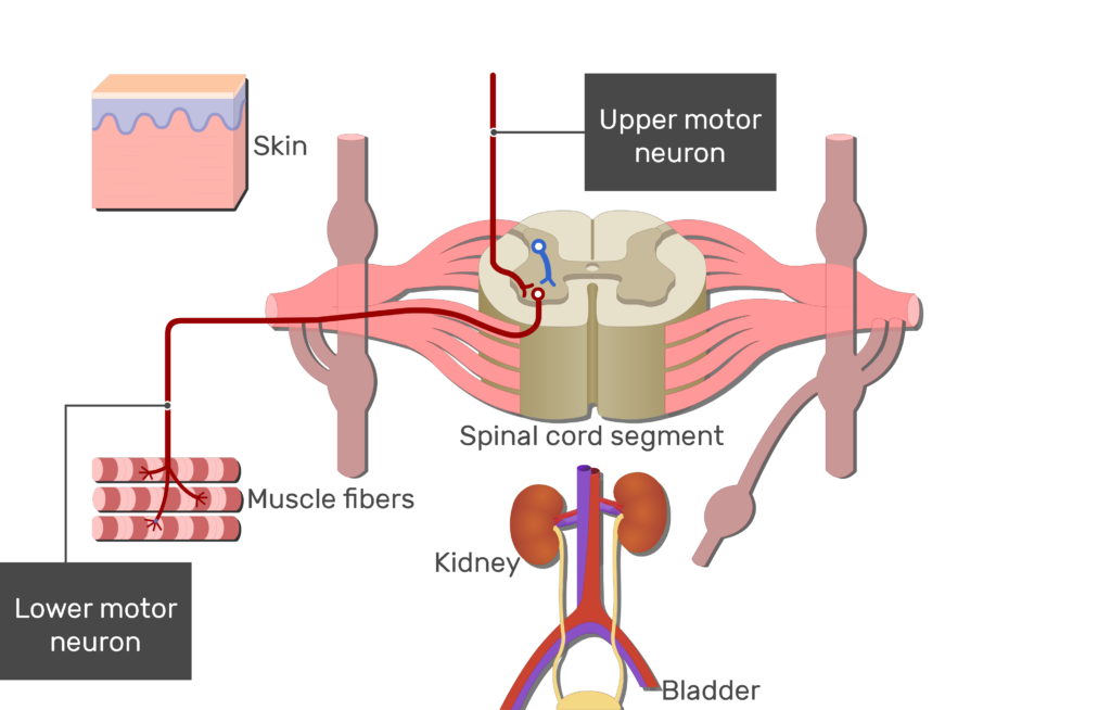
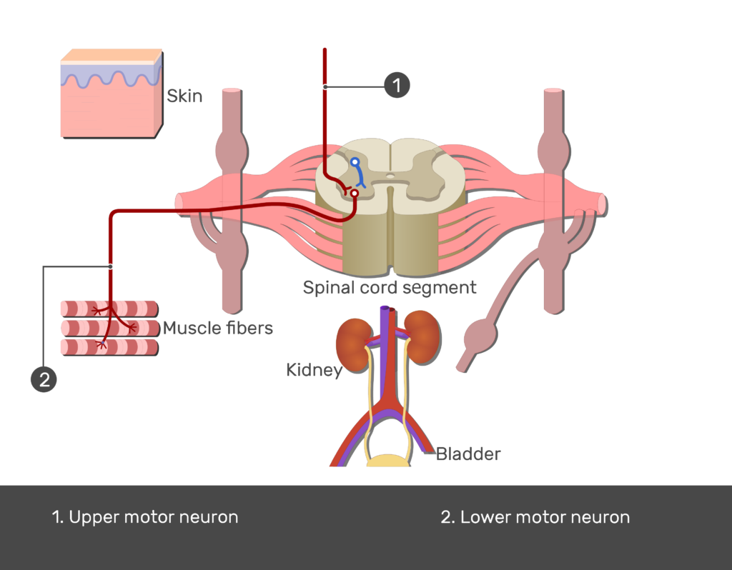
- In the ventral horns (or anterior horns), descending upper level motor neurons (and other types of neurons) synapse with outgoing lower level motor neurons.
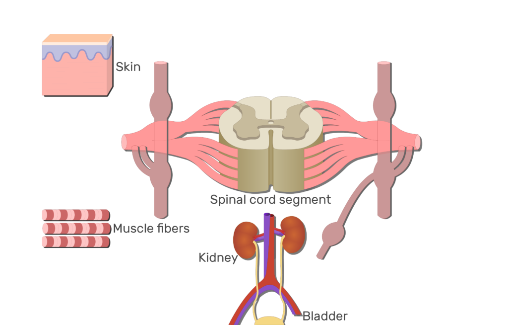
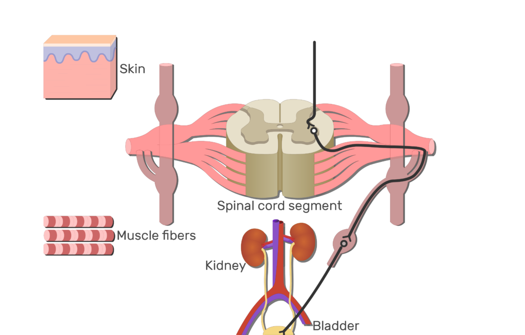
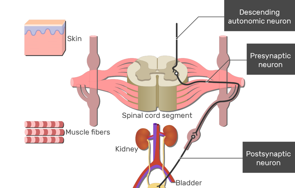
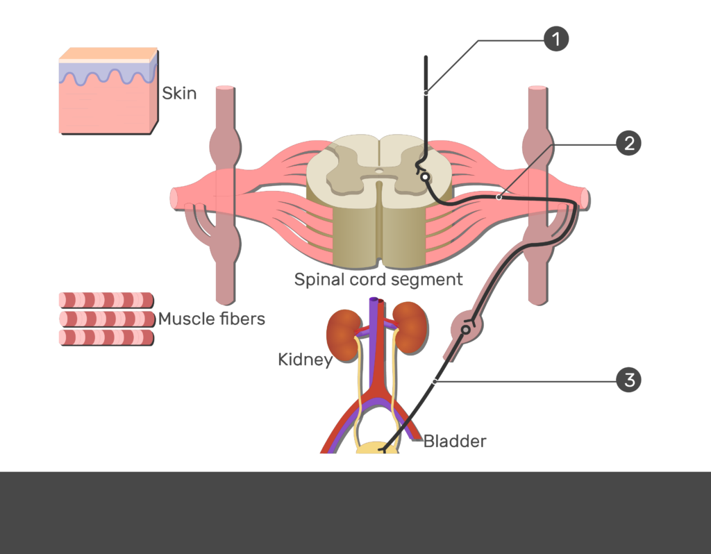
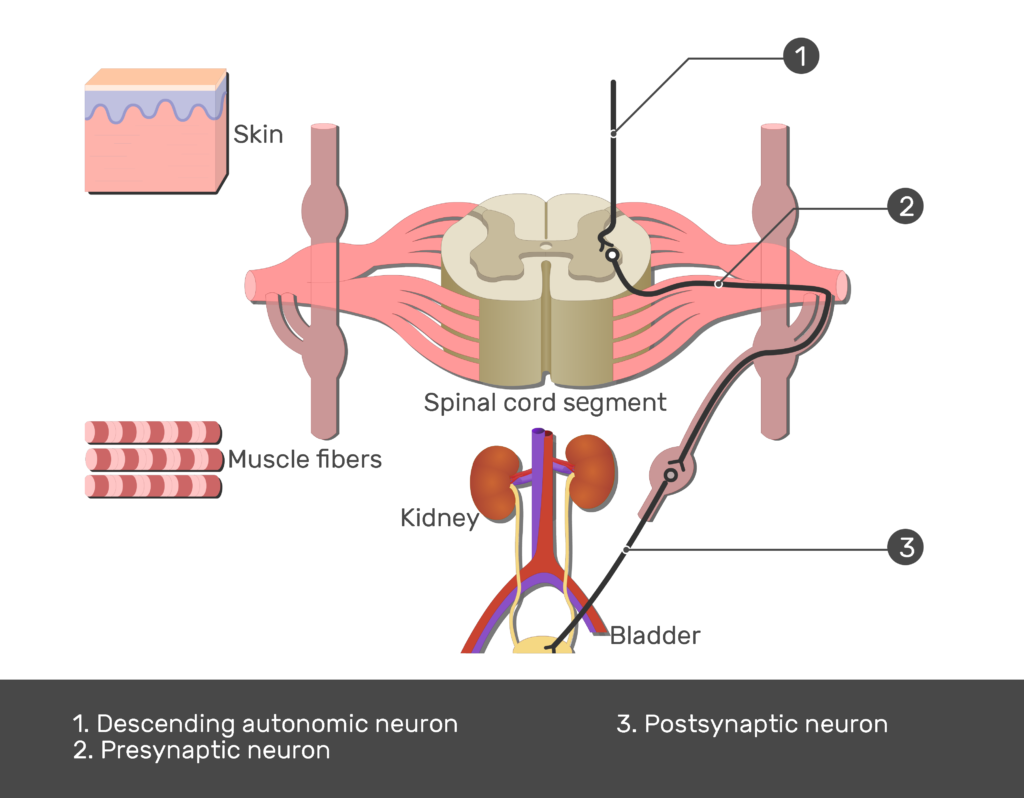
- In the lateral horns, descending autonomic pathway neurons synapse with outgoing sympathetic motor neurons.
Pathways of the spinal cord gray matter horns.
Overview:
[ ![]() Dorsal horn /
Dorsal horn / ![]() Ventral horn /
Ventral horn / ![]() Lateral horn ]
Lateral horn ]


Related Articles

Spinal Nerve Roots
The spinal nerve roots are two pairs extend from each segment of the spinal cord which explained beautifully in an illustrated and interactive way.
Spinal Cord White Matter – Anatomy & Functions
Spinal Cord White Matter (Anatomy & Functions); explained beautifully in an illustrated and interactive way.
Spinal Cord Segments – Cross-sectional Anatomy
The spinal cord is made up of 31 segments, this tutorial shows some anatomy, cross section and histology images of the segments in interactive way.