


Trachea: anatomy, structure and function
Bronchus and bronchial wall: anatomy and function
Last update:
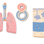
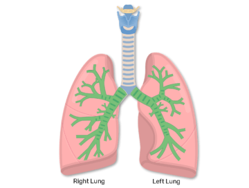
The bronchi are part of the airway system of the lower respiratory system. Bronchi are the branches of the trachea which provide oxygen to the lungs.
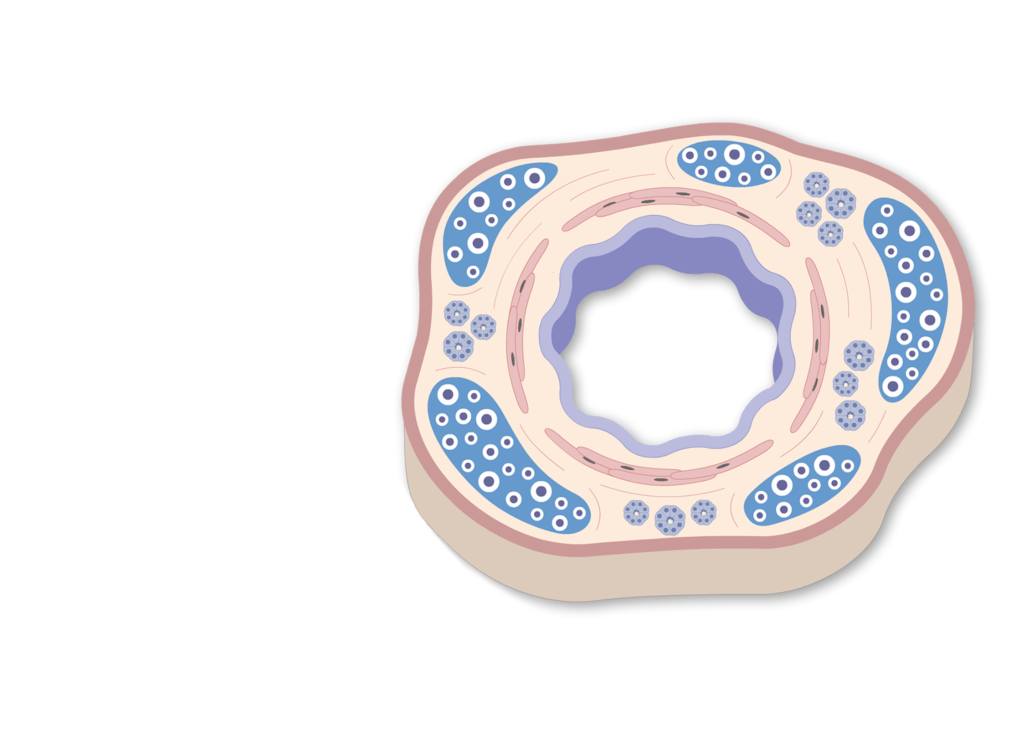
- In cross-section, the
 bronchial wall appears similar to the trachea.
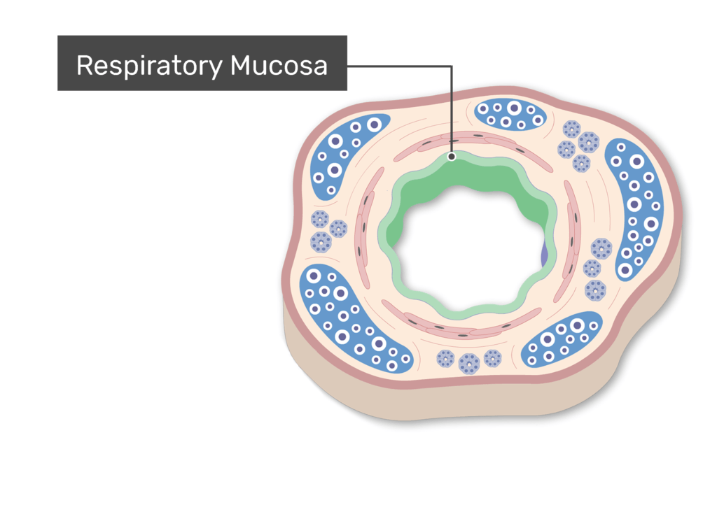
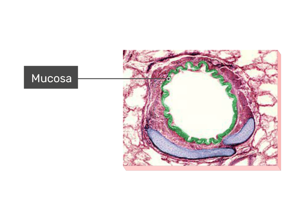
- Respiratory mucosa (or mucous membrane) lines the luminal surface. Mucus-secreting goblet cells are present in the epithelium. However, they are less numerous than in the trachea.
- Deep to the mucosa is a layer containing smooth muscle fibers, hyaline cartilage, and scattered seromucous and mucous glands.
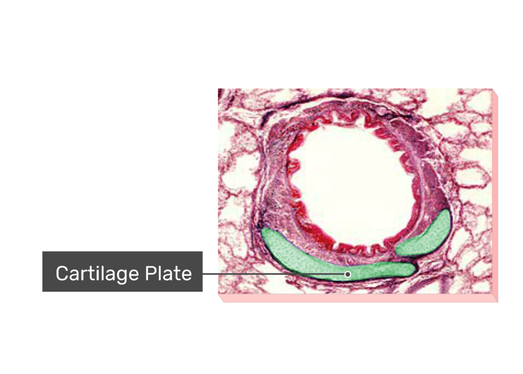
- The cartilage appears as rings in the larger bronchi but changes to irregular-sized plates in the smaller bronchi. As in the trachea, the cartilage helps keep the bronchial wall from collapsing.
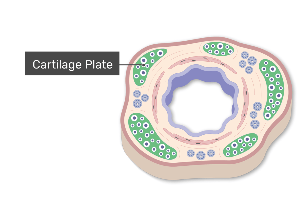
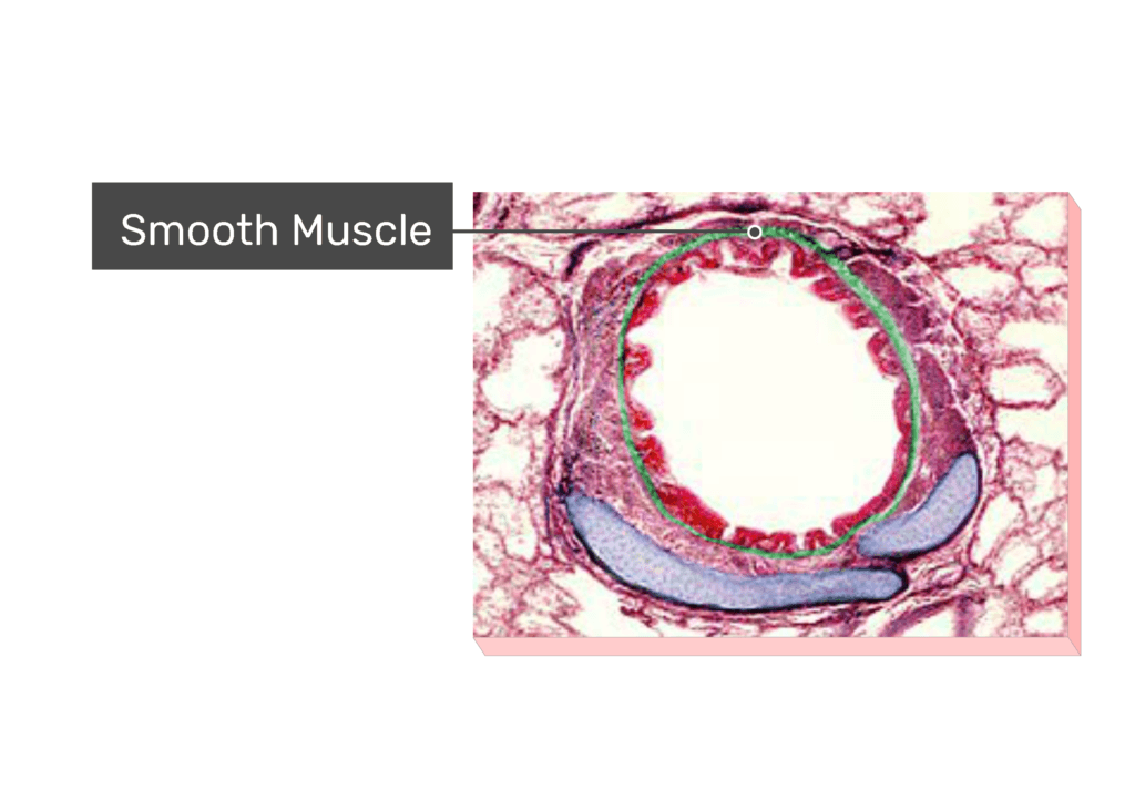
- The smooth muscle fibers are located between the mucosa and cartilage plates and form a nearly complete ring.
Master respiratory system anatomy with these interactive quizzes and labeling exercises.

- They are involuntarily controlled and their
 movement alters the size of the bronchial lumen. The changes in lumen size may increase airflow during normal breathing, protect lungs tissues from foreign particles and irritants, or improve the effectiveness of a cough.
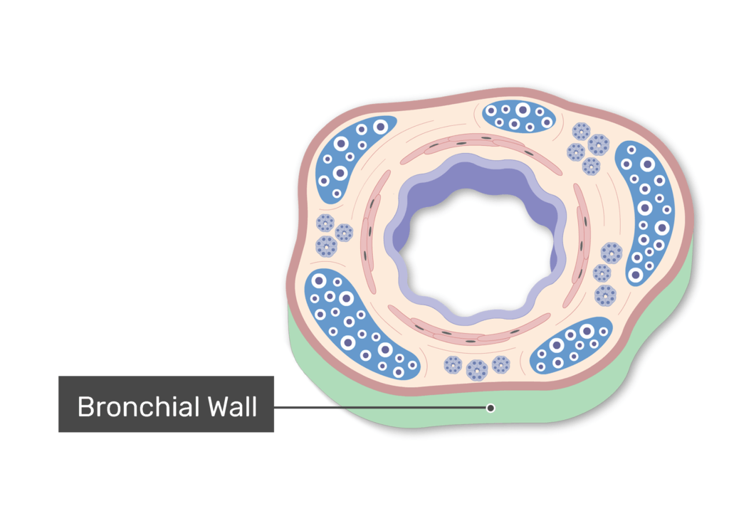
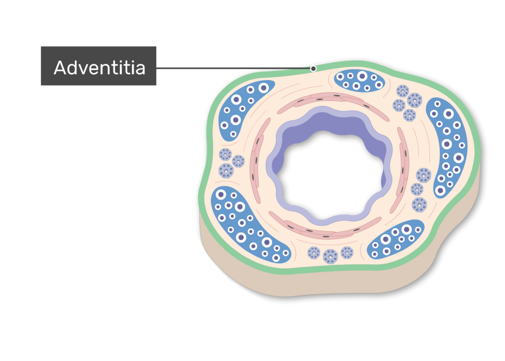
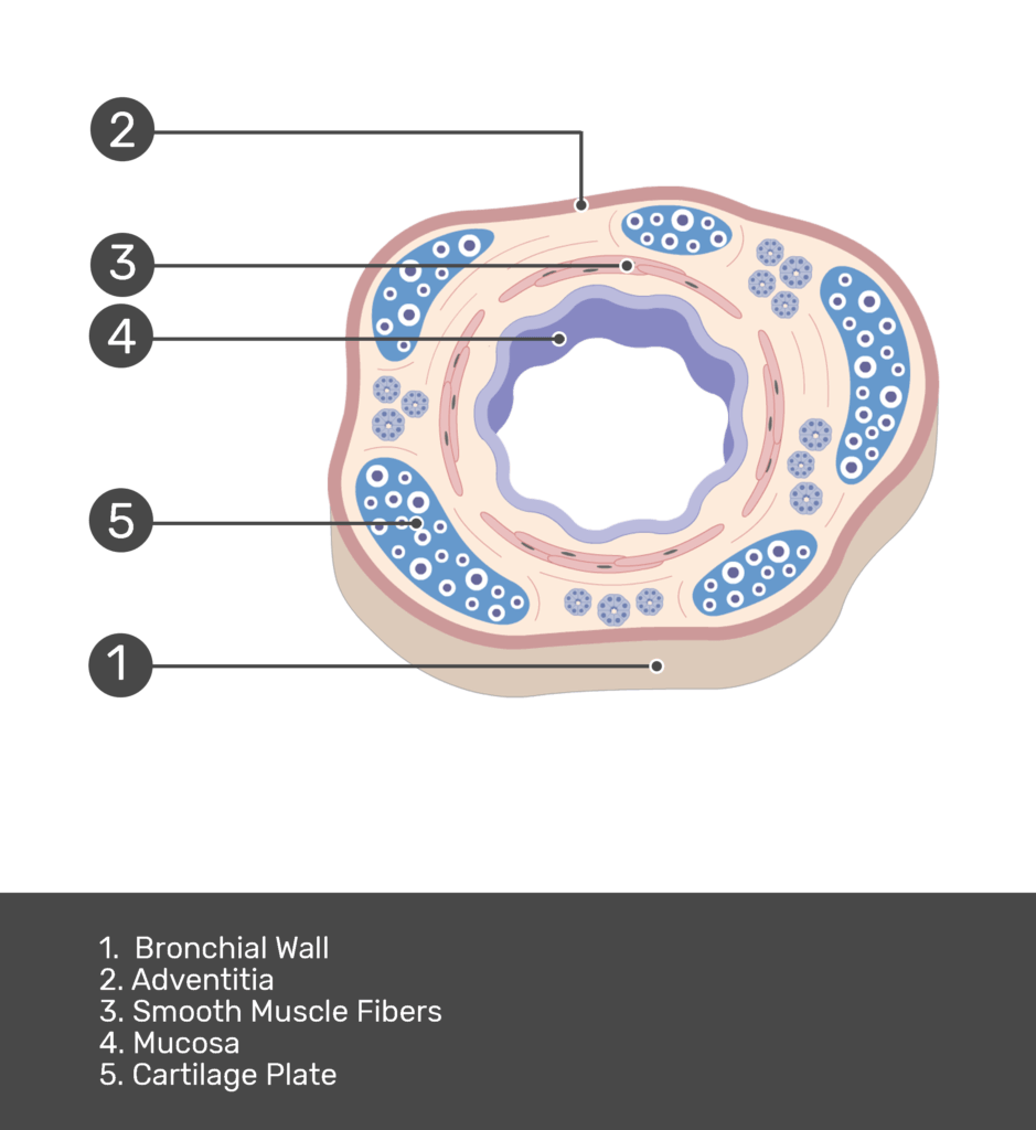
- A narrow band of adventitia covers the outer bronchial wall, which connects the bronchus to the surrounding lung tissues.
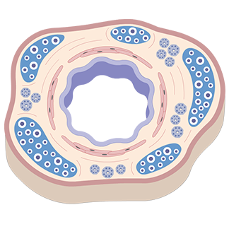
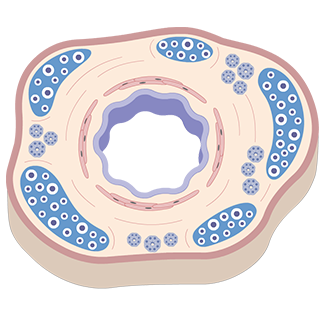
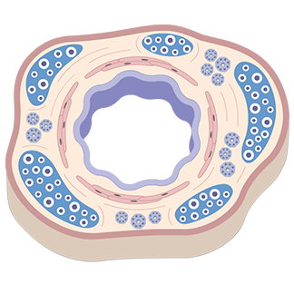
Photomicrograph of the bronchial wall:
Components: ![]() Cartilage plate,
Cartilage plate, ![]() mucosa,
mucosa, ![]() smooth muscle &
smooth muscle & ![]() lung tissue
lung tissue


An Overview of the Bronchial Wall Anatomy:

Test yourself while observing the Bronchial Wall Structure
Related Articles
Trachea: anatomy, structure and function
There are four tissue layers of the tracheal wall. The trachea includes respiratory mucosa, submucosa, cartilaginous rings, trachealis muscle, and adventitia.
Bronchial Tubes Structure, Functions, & Location | Bronchus Anatomy
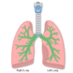
Near the sternal angle, the trachea bifurcates (or splits), into the right and left primary (1) bronchi. Each bronchus runs freely for a few centimeters, then enters its respective lung. Air flows in and out of each lung through the primary bronchi.