Anatomy of the Hand and Wrist Bones
Carpal Bones
Last update:
The carpal bones are eight irregular bones that form the root of the hand. They form the region known as carpus, often frequently termed as simply the wrist.
The carpal bones are distributed into two rows, each consisting of four bones:
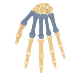
- Proximal row: Scaphoid bone, lunate bone, triquetrum bone, pisiform bone
- Distal row: Trapezium bone, trapezoid bone, capitate bone, hamate bone
The proximal row of the carpal bones articulate with the radius to form the wrist joint (radiocarpal joint). The distal row articulates with the metacarpal bones, forming five carpometacarpal (CMC) joints. The proximal and distal rows glide against each other, ensuring the proper synchronization of the movements which eventually result with the proper mobility of the hand.
This article will describe the anatomy of the carpal bones.
| Key points about the carpal bones | |
|---|---|
| Definition and location | A set of 8 irregular bones located at the root of the palm. |
| Proximal row | Scaphoid Lunate Triquetrum Pisiform |
| Distal row | Trapezium Trapezoid Capitate Hamate |
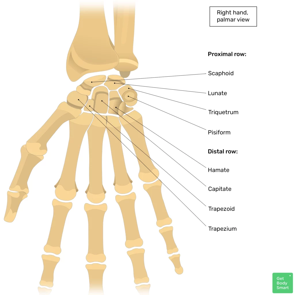
Carpal bones: Labeled diagram
Proximal row
From lateral to medial, the proximal carpal bones are:
- Scaphoid bone is the largest bone of the proximal row. On its proximal surface, the scaphoid contains an articular facet that participates in the joint with the radius (wrist/radiocarpal joint).
- Lunate bone is a moon-shaped bone, which also contributes to the wrist joint.
- Triquetrum bone is a pyramidal shaped bone, and the final of the three carpals that participate in the wrist joint.
- Pisiform bone is a pea shaped bone that is located on the anterior (palmar) surface of the triquetrum bone. Thus, it is not visible from the dorsal (posterior) perspective of the hand.
Use these interactive carpal bones quizzes to learn and remember the carpal bones for good!
Distal row
From lateral to media, the distal carpal bones are:
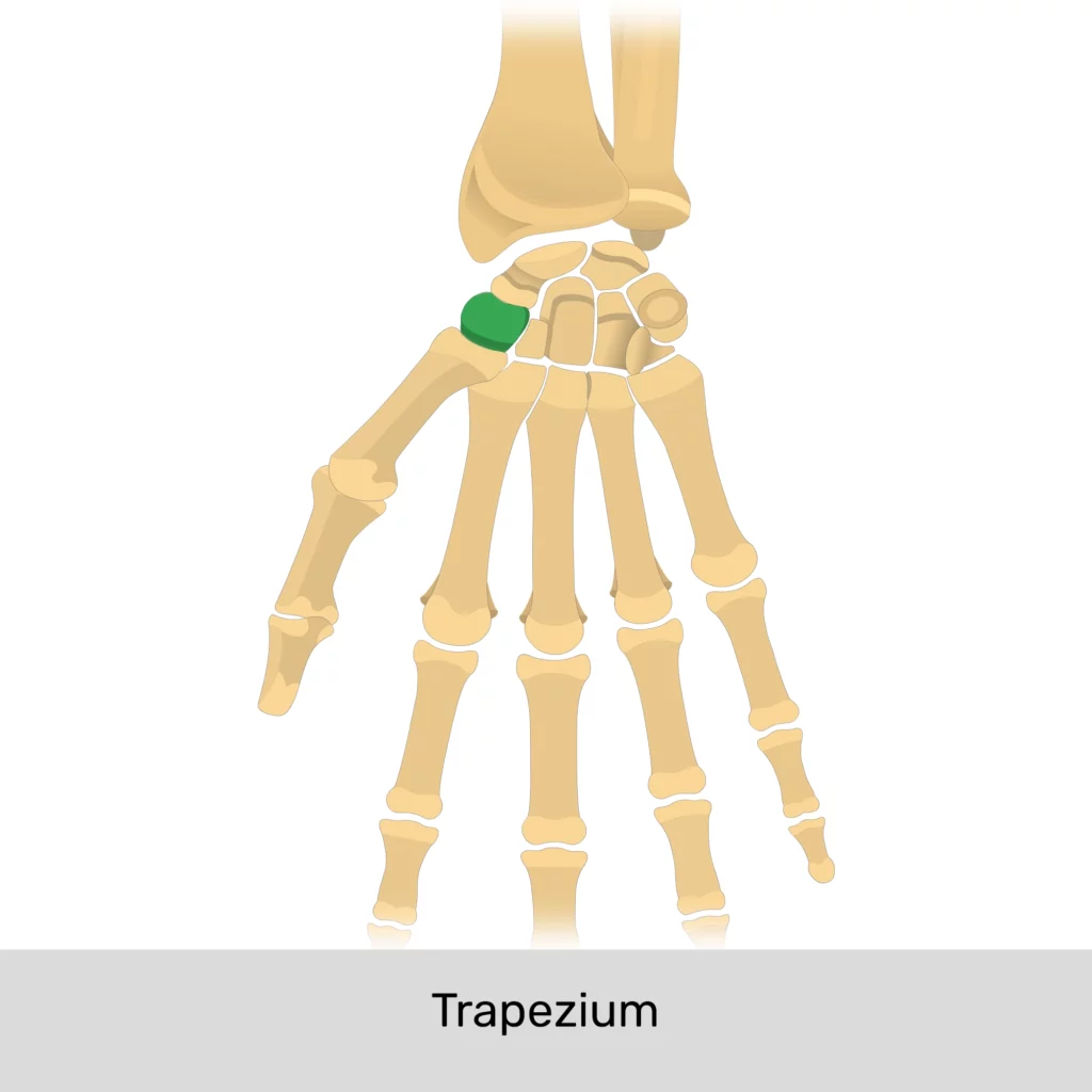
- Trapezium bone is a quadrangular bone. It articulates with the first metacarpal bone, which is the metacarpal of the thumb. This carpometacarpal (CMC) joint is also known as the trapeziometacarpal joint and it is the most mobile CMC joint of all.
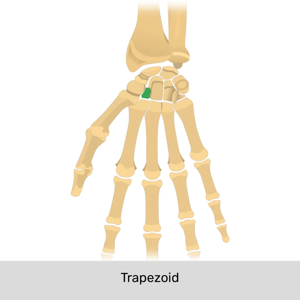
- Trapezoid bone is wedge-shaped and significantly smaller than the trapezium bone. It articulates with the second metacarpal (index finger), as well as the trapezium, capitate and scaphoid bones.
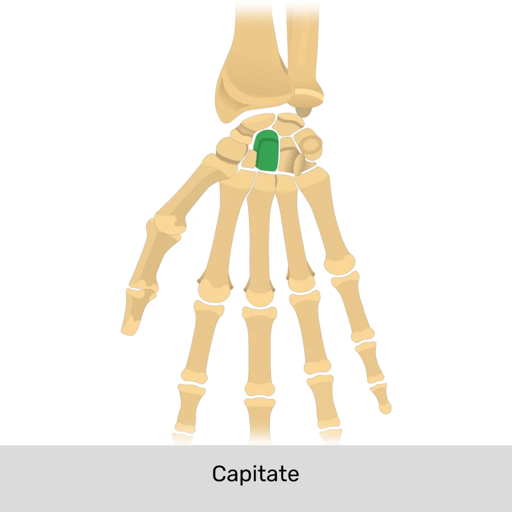
- Capitate bone is the largest carpal bone. It forms joints with the third metacarpal (middle finger), trapezoid, scaphoid, lunate and hamate bones.
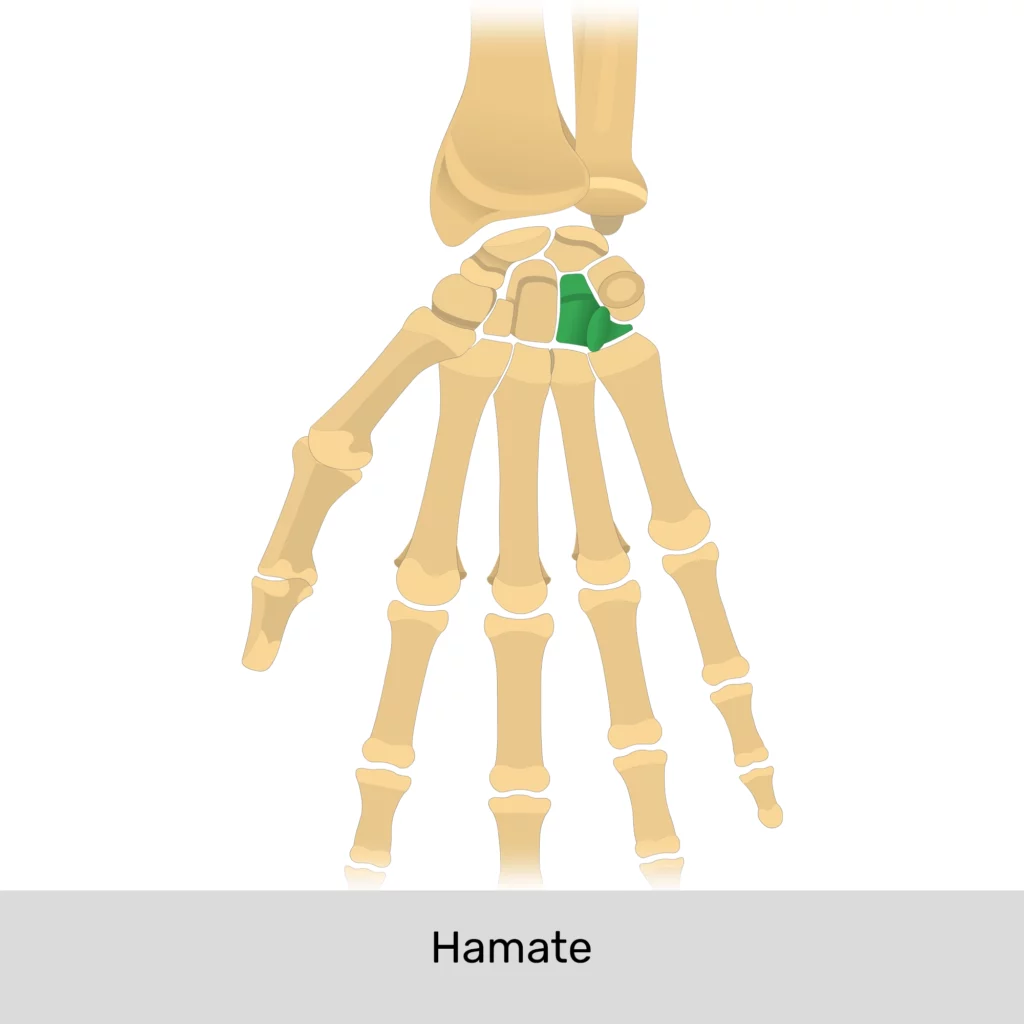
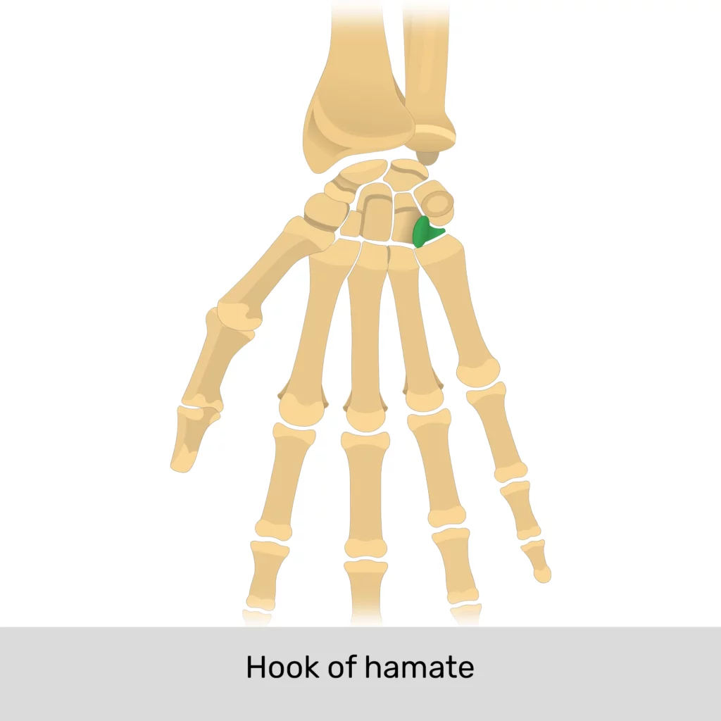
- Hamate bone forms two CMC joints: with the fourth and fifth metacarpals (ring and pinky fingers). Its distinctive feature is the hook of the hamate bone, visible on its anterior surface.
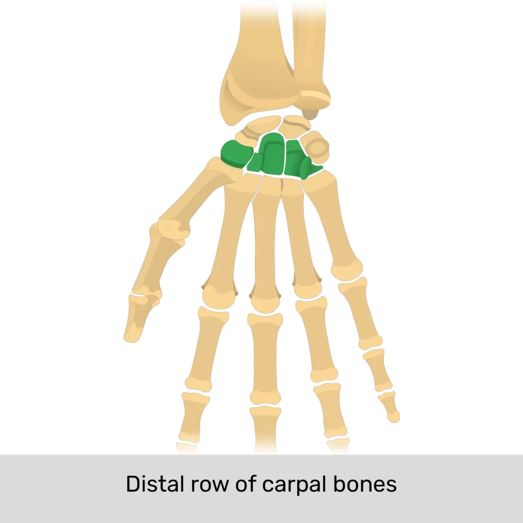
Distal row of the carpal bones
Mnemonic
A useful mnemonic for remembering the names of the carpal bones is “She Likes To Play Try To Catch Her”:
| She | Scaphoid |
| Likes | Lunate |
| To | Triquetrum |
| Play | Pisiform |
| Try | Trapezium |
| To | Trapezoid |
| Catch | Capitate |
| Her | Hamate |
Carpal bones quiz
The following quiz will help you solidify your knowledge about the carpal bones:
References
- Open Anatomy. (n.d.). TA2 Viewer. Retrieved April 5, 2023, from https://ta2viewer.openanatomy.org/
- Moore, K. L. (2018). Clinically Oriented Anatomy (8th ed.). Philadelphia, PA: Wolters Kluwer.
- Drake, R. L., Vogl, A. W., & Mitchell, A. W. M. (2015). Gray’s Anatomy for Students (3rd ed.). Edinburgh, Scotland: Churchill Livingstone.
- Standring, S. (2021). Gray’s Anatomy (42tst ed.). Edinburgh: Elsevier Churchill Livingstone.
Related Articles
Scapula Bone Quiz
This 2-part quiz tests your knowledge on the anatomical markings of […]
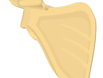
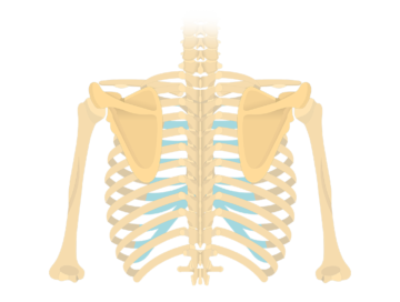
Scapula Bone
The bone markings of the anterior scapula include the superior border, medial border, lateral border, superior angle, lateral angle, inferior angle, coracoid process, suprascapular notch, glenoid cavity, infraglenoid tubercle, and the suprascapular fossa.
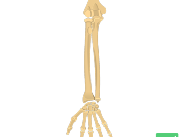
Radius and Ulna Bones Anatomy
An interactive tutorial covering the anatomy of the radius and ulna, the two bones that comprises the forearm.