Clavicle Bone Anatomy
Scapula Bone
Last update:
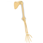
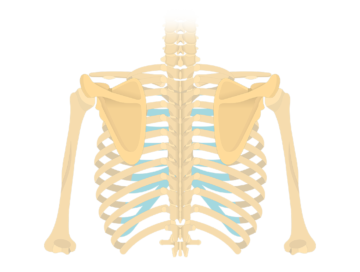
The scapula or shoulder blade is a flat, triangular upper limb bone that lies on the posterior surface of the thorax, over the ribs 2-7. It is a part of the pectoral (shoulder) girdle, together with the clavicle.
The scapula articulates with the humerus, forming the shoulder (glenohumeral) joint. It also articulates with the clavicle via the acromioclavicular (AC) joint. This serial connection between these bones gives the scapula an important role in the mobility of the upper limb. Primarily, it supports the shoulder joint and helps in transmission of force from the upper limb to the trunk, ensuring the proper joint functioning. In addition, it provides attachment to a number of shoulder muscles.
This article will discuss the anatomical landmarks of the scapula, as well as the muscles that attach to it.
| Key points about the anatomy of the scapula | |
|---|---|
| Surfaces | Anterior (costal): subscapular fossa Posterior (dorsal): spine of scapula, supraspinous fossa, infraspinous fossa, acromion |
| Angles | Lateral: glenoid process, neck of scapula Superior Inferior |
| Borders (margins) | Superior, lateral, medial |
| Muscles that attach to the scapula | Originate: deltoid, supraspinatus, infraspinatus, triceps brachii (long head), teres minor, teres major, latissimus dorsi, coracobrachialis, biceps brachii (long and short heads), subscapularis, omohyoid Insert: trapezius, levator scapulae, rhomboid major, rhomboid minor, serratus anterior, pectoralis minor |
Anatomy
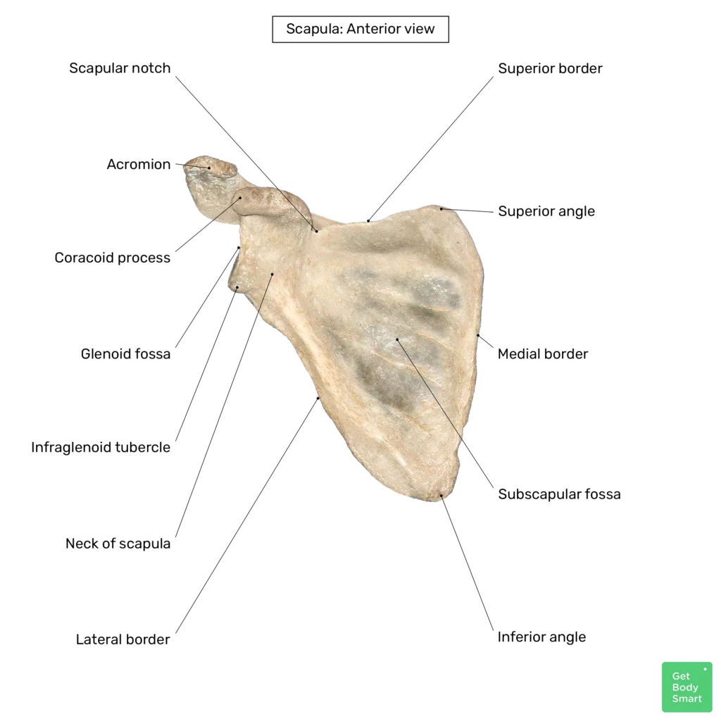
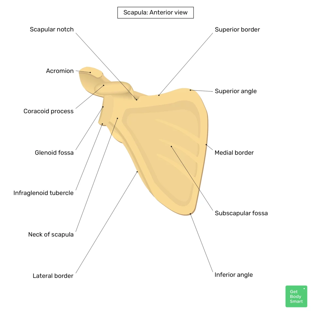
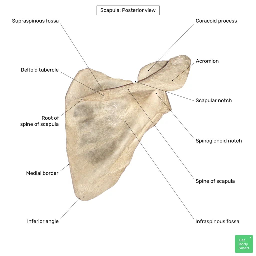
The scapula has three borders (superior, medial and lateral) and three angles (superior, inferior and lateral). It has two surfaces, namely the anterior (costal) surface and posterior surface. The scapula also features a prominent projection that arises from its superior surface, called the acromion.

Landmarks of the scapula
Surfaces
The anterior (costal) surface of the scapula is mostly occupied by a large, slightly depressed region called the subscapular fossa. This fossa serves as an attachment for the subscapularis muscle. The posterior (dorsal) surface features a prominent ridge called the spine of scapula, which divides the surface into two unequal parts called the supraspinous fossa and infraspinous fossa.
Keep learning the anatomy of the scapula with quizzes and labeled diagrams.
The spine crosses the dorsal surface in a superiolateral direction. Near its root, the spine features a notable bump called the deltoid tubercle, which serves as an attachment for the deltoid muscle. The spine terminates with a lateral projection called the acromion. The acromion features the articular surface called the clavicular facet, via which it participates in the joint with the clavicle (acromioclavicular joint). Since the acromion emerges from the dorsal surface of the scapula, it creates a small notch with the dorsal surface called the spinoglenoid notch.
Angles
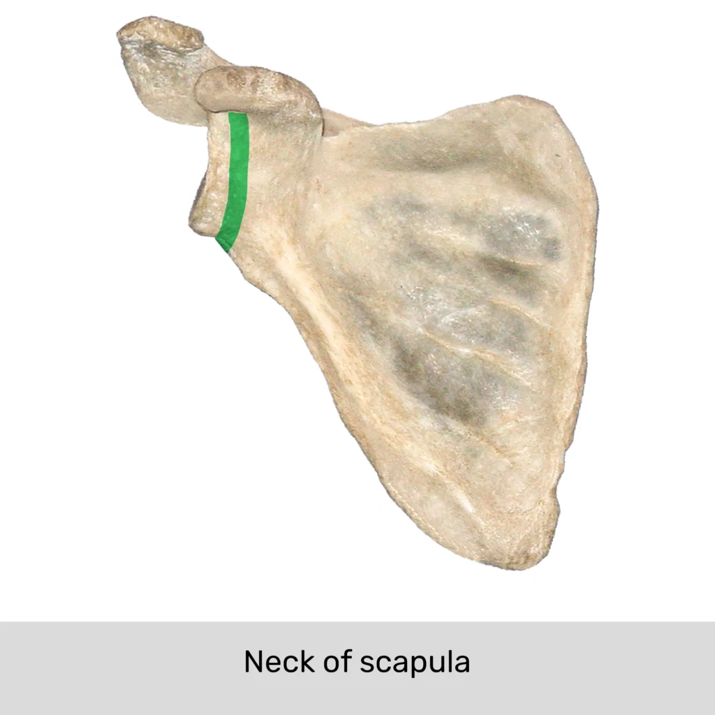
Scapula has three angles, lateral, superior and inferior. The superior and inferior angles of the scapula are unremarkable. The lateral angle features a region called the glenoid process of scapula. The glenoid process is connected to the lateral angle via a constriction called the neck of scapula.
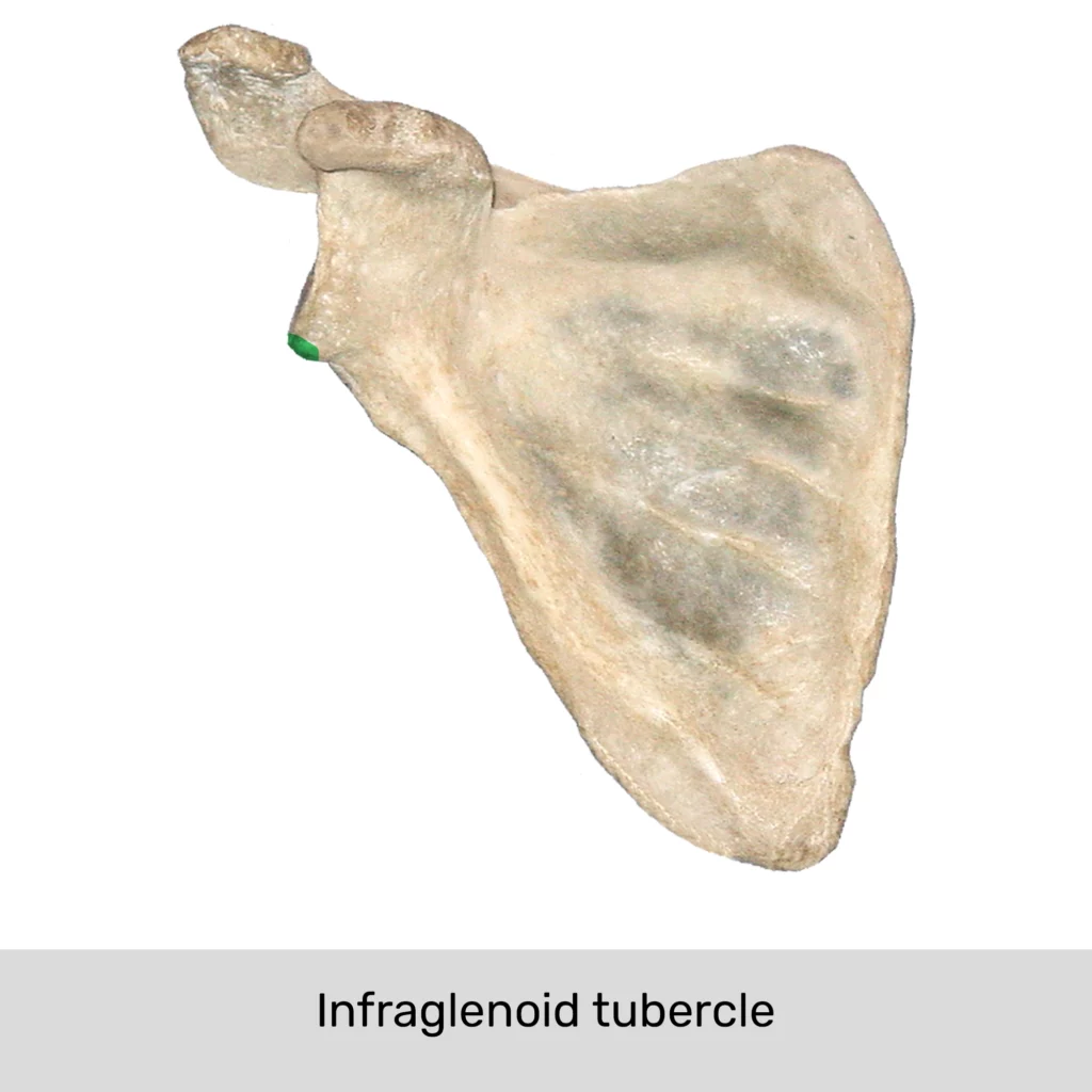
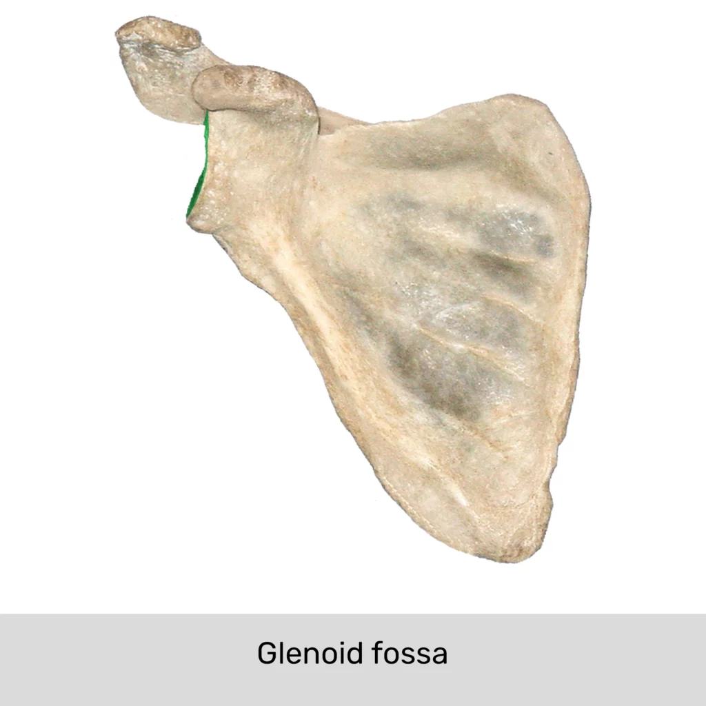
The glenoid process features an articular surface called the glenoid fossa, via which it articulates with the head of the humerus and forms the glenohumeral (shoulder) joint. The superior and inferior rims of the fossa feature supraglenoid and infraglenoid tubercles, respectively.
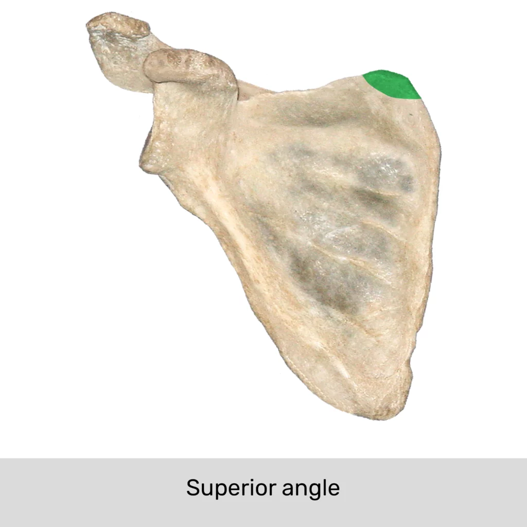

Angles of the scapula and their associated landmarks.
Borders
The scapula has superior, lateral and medial borders. Only the superior border contains notable bony markings: the scapular notch and coracoid process. The scapular notch is a noticeable dip in the superior border. It is closed by the suprascapular ligament, and therefore transformed into the scapular canal. This canal is traversed by the suprascapular nerve and vessels.
Laterally to the notch, there is a bony projection directed anteriorly and laterally called the coracoid process. Together with the acromion, this process serves to stabilize the shoulder joint.
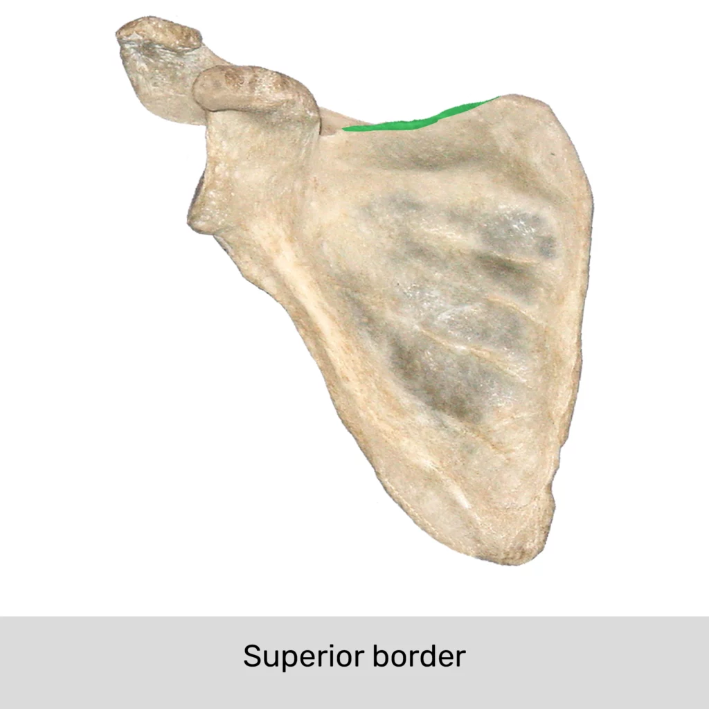
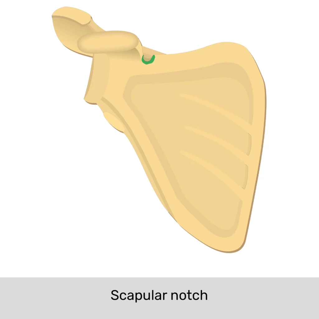
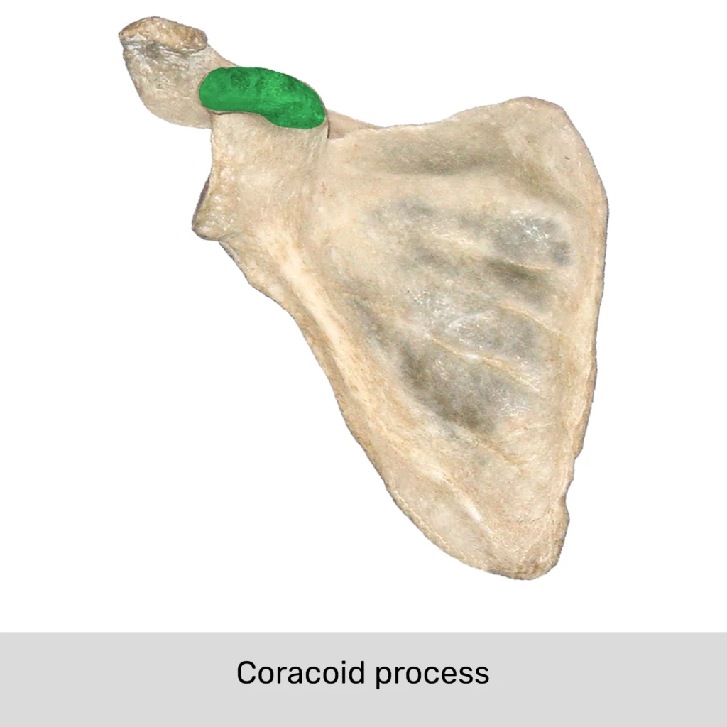
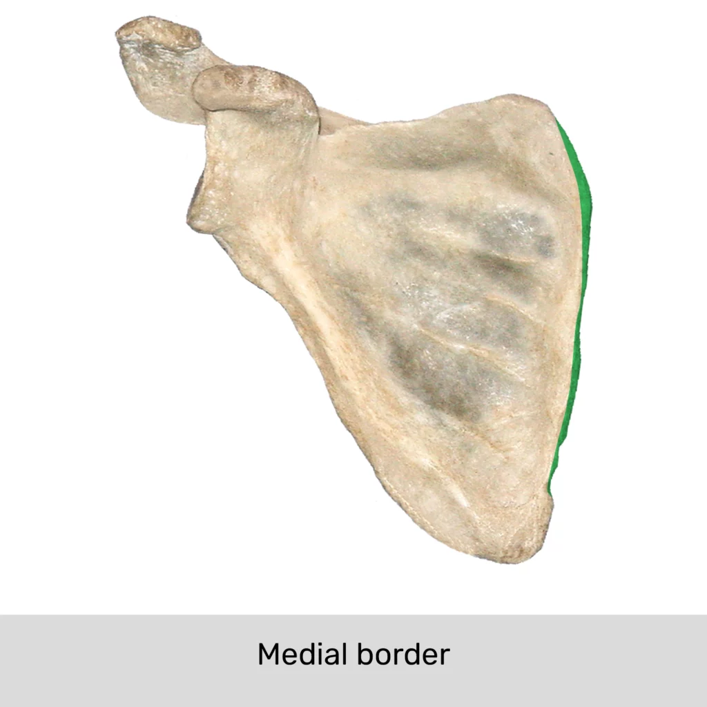
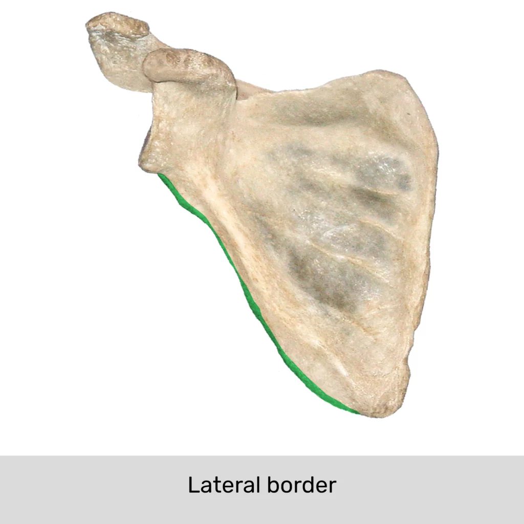
Borders of the scapula and their landmarks.
Muscles that attach to scapula
There are 17 muscles in total that attach to the scapula. These include the muscles that stabilize the shoulder joint (rotator cuff muscles), as well as those whose primary function is to provide it with movement. Namely, they are:
- Muscles that originate from the scapula: deltoid, supraspinatus, infraspinatus, triceps brachii (long head), teres minor, teres major, latissimus dorsi, coracobrachialis, biceps brachii (long and short heads), subscapularis, omohyoid
- Muscles that insert to scapula: trapezius, levator scapulae, rhomboid major, rhomboid minor, serratus anterior and pectoralis minor.
The following diagram shows all the muscles that originate from and insert to scapula:
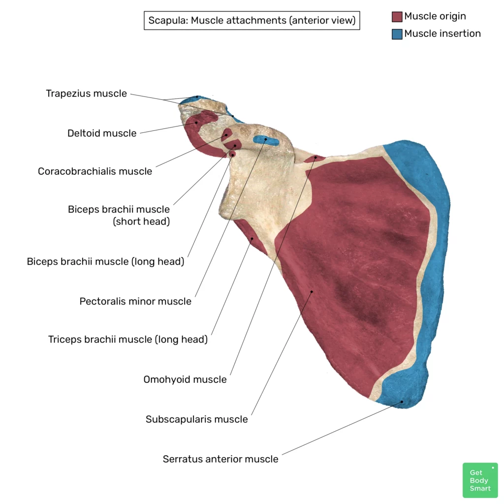
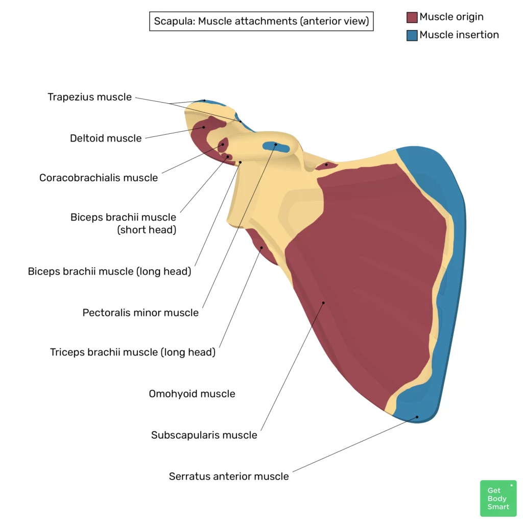

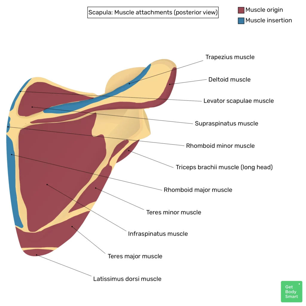
Muscles of the scapula
Scapula anatomy quiz
The following quiz will help you solidify your knowledge about the bony markings of the scapula, as well as the muscles that attach to the scapula:
References
- Open Anatomy. (n.d.). TA2 Viewer. Retrieved April 5, 2023, from https://ta2viewer.openanatomy.org/
- Moore, K. L. (2018). Clinically Oriented Anatomy (8th ed.). Philadelphia, PA: Wolters Kluwer.
- Drake, R. L., Vogl, A. W., & Mitchell, A. W. M. (2015). Gray’s Anatomy for Students (3rd ed.). Edinburgh, Scotland: Churchill Livingstone.
- Standring, S. (2021). Gray’s Anatomy (42tst ed.). Edinburgh: Elsevier Churchill Livingstone.
Related Articles
Scapula Bone Quiz
This 2-part quiz tests your knowledge on the anatomical markings of […]
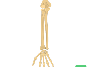
Radius and Ulna Bones Anatomy
An interactive tutorial covering the anatomy of the radius and ulna, the two bones that comprises the forearm.
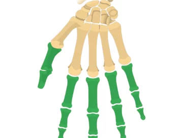
Phalanges of the hand (phalanx bones)
Each finger consists of three phalanges: Proximal phalanges, middle phalanges, and distal phalanges The thumb only has two: Proximal phalanges and distal phalanges.